5UUY | pdb_00005uuy

Crystal structure of Dioclea lasiocarpa lectin (DLL) complexed with X-MAN


Experimental Data Snapshot

  • Method: X-RAY DIFFRACTION
  • Resolution: 1.88 Å
  • R-Value Free: 
    0.262 (Depositor), 0.271 (DCC) 
  • R-Value Work: 
    0.225 (Depositor) 
  • R-Value Observed: 
    0.227 (Depositor) 

Starting Model: experimental
View more details

wwPDB Validation 3D Report Full Report

Validation slider image for 5UUY

Ligand Structure Quality Assessment 


This is version 1.4 of the entry. See complete history

Literature

Structural analysis of Dioclea lasiocarpa lectin: A C6 cells apoptosis-inducing protein.

Nascimento, K.S.Santiago, M.Q.Pinto-Junior, V.R.Osterne, V.J.S.Martins, F.W.V.Nascimento, A.P.M.Wolin, I.A.V.Heinrich, I.A.Martins, M.G.Q.Silva, M.T.L.Lossio, C.F.Rocha, C.R.C.Leal, R.B.Cavada, B.S.

(2017) Int J Biochem Cell Biol 92: 79-89

  • DOI: https://doi.org/10.1016/j.biocel.2017.09.014
  • Primary Citation Related Structures: 
    5UUY

  • PubMed Abstract: 

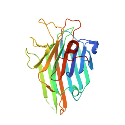
    Lectins are multidomain proteins that specifically recognize various carbohydrates. The structural characterization of these molecules is crucial in understanding their function and activity in systems and organisms. Most cancer cells exhibit changes in glycosylation patterns, and lectins may be able to recognize these changes. In this work, Dioclea lasiocarpa seed lectin (DLL) was structurally characterized. The lectin presented a high degree of similarity with other lectins isolated from legumes, presenting a jelly roll motif and a metal-binding site stabilizing the carbohydrate-recognition domain. DLL demonstrated differential interactions with carbohydrates, depending on type of glycosidic linkage present in ligands. As observed by the reduction of cell viability in C6 cells, DLL showed strong antiglioma activity by mechanisms involving activation of caspase 3.


  • Organizational Affiliation
    • Universidade Federal do Ceara (UFC), Fortaleza, Ceara, Brazil.

Macromolecule Content 

  • Total Structure Weight: 25.94 kDa 
  • Atom Count: 1,923 
  • Modeled Residue Count: 231 
  • Deposited Residue Count: 237 
  • Unique protein chains: 1

Macromolecules

Find similar proteins by:|  3D Structure
Entity ID: 1
MoleculeChains  Sequence LengthOrganismDetailsImage
Dioclea lasiocarpa lectin (DLL)237DiocleaMutation(s): 0 
UniProt
Find proteins for P58907 (Dioclea virgata)
Explore P58907 
Go to UniProtKB:  P58907
Entity Groups
Sequence Clusters30% Identity50% Identity70% Identity90% Identity95% Identity100% Identity
UniProt GroupP58907
Sequence Annotations
Expand
Reference Sequence

Experimental Data & Validation

Experimental Data

  • Method: X-RAY DIFFRACTION
  • Resolution: 1.88 Å
  • R-Value Free:  0.262 (Depositor), 0.271 (DCC) 
  • R-Value Work:  0.225 (Depositor) 
  • R-Value Observed: 0.227 (Depositor) 
Space Group: I 2 2 2
Unit Cell:
Length ( Å )Angle ( ˚ )
a = 64.66α = 90
b = 85.98β = 90
c = 93.25γ = 90
Software Package:
Software NamePurpose
SCALAdata scaling
PHENIXrefinement
PDB_EXTRACTdata extraction
MOLREPphasing
iMOSFLMdata reduction

Structure Validation

View Full Validation Report


Ligand Structure Quality Assessment 


Entry History 

Deposition Data

Revision History  (Full details and data files)

  • Version 1.0: 2017-10-04
    Type: Initial release
  • Version 1.1: 2017-10-11
    Changes: Database references
  • Version 1.2: 2018-04-18
    Changes: Data collection
  • Version 1.3: 2020-07-29
    Type: Remediation
    Reason: Carbohydrate remediation
    Changes: Derived calculations, Structure summary
  • Version 1.4: 2023-10-04
    Changes: Data collection, Database references, Refinement description, Structure summary