5ULW | pdb_00005ulw

Structure of human DNA polymerase iota bound to template 1-methyl-deoxyadenosine


Experimental Data Snapshot

  • Method: X-RAY DIFFRACTION
  • Resolution: 2.62 Å
  • R-Value Free: 
    0.248 (Depositor), 0.248 (DCC) 
  • R-Value Work: 
    0.204 (Depositor), 0.207 (DCC) 
  • R-Value Observed: 
    0.207 (Depositor) 

Starting Model: experimental
View more details

wwPDB Validation 3D Report Full Report

Validation slider image for 5ULW

Ligand Structure Quality Assessment 


This is version 1.3 of the entry. See complete history

Literature

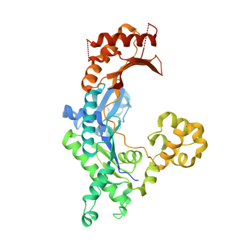
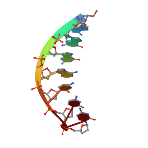
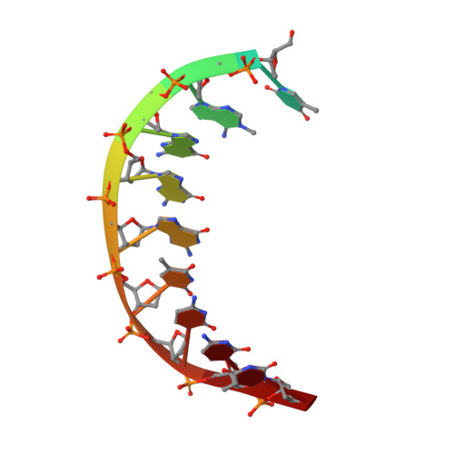
Mechanism of error-free DNA synthesis across N1-methyl-deoxyadenosine by human DNA polymerase-iota.

Jain, R.Choudhury, J.R.Buku, A.Johnson, R.E.Prakash, L.Prakash, S.Aggarwal, A.K.

(2017) Sci Rep 7: 43904-43904

  • DOI: https://doi.org/10.1038/srep43904
  • Primary Citation Related Structures: 
    5ULW, 5ULX

  • PubMed Abstract: 

    N1-methyl-deoxyadenosine (1-MeA) is formed by methylation of deoxyadenosine at the N1 atom. 1-MeA presents a block to replicative DNA polymerases due to its inability to participate in Watson-Crick (W-C) base pairing. Here we determine how human DNA polymerase-ι (Polι) promotes error-free replication across 1-MeA. Steady state kinetic analyses indicate that Polι is ~100 fold more efficient in incorporating the correct nucleotide T versus the incorrect nucleotide C opposite 1-MeA. To understand the basis of this selectivity, we determined ternary structures of Polι bound to template 1-MeA and incoming dTTP or dCTP. In both structures, template 1-MeA rotates to the syn conformation but pairs differently with dTTP versus dCTP. Thus, whereas dTTP partakes in stable Hoogsteen base pairing with 1-MeA, dCTP fails to gain a "foothold" and is largely disordered. Together, our kinetic and structural studies show how Polι maintains discrimination between correct and incorrect incoming nucleotide opposite 1-MeA in preserving genome integrity.


  • Organizational Affiliation
    • Department of Pharmacological Sciences, Icahn School of Medicine at Mount Sinai, Box 1677, 1425 Madison Avenue, New York, NY 10029, USA.

Macromolecule Content 

  • Total Structure Weight: 53.02 kDa 
  • Atom Count: 3,224 
  • Modeled Residue Count: 389 
  • Deposited Residue Count: 438 
  • Unique protein chains: 1
  • Unique nucleic acid chains: 2

Macromolecules


Find similar proteins by:|  3D Structure
Entity ID: 3
MoleculeChains  Sequence LengthOrganismDetailsImage
DNA polymerase iotaC [auth A]420Homo sapiensMutation(s): 0 
Gene Names: POLIRAD30B
EC: 2.7.7.7
UniProt & NIH Common Fund Data Resources
Find proteins for Q9UNA4 (Homo sapiens)
Explore Q9UNA4 
Go to UniProtKB:  Q9UNA4
PHAROS:  Q9UNA4
GTEx:  ENSG00000101751 
Entity Groups
Sequence Clusters30% Identity50% Identity70% Identity90% Identity95% Identity100% Identity
UniProt GroupQ9UNA4
Sequence Annotations
Expand
Reference Sequence
Find similar nucleic acids by:  Sequence
Entity ID: 1
MoleculeChains LengthOrganismImage
DNA (5'-D(*AP*GP*GP*AP*CP*CP*(DOC))-3')A [auth P]7unidentified
Sequence Annotations
Expand
Reference Sequence
Find similar nucleic acids by:  Sequence
Entity ID: 2
MoleculeChains LengthOrganismImage
DNA (5'-D(*TP*(MA7)P*GP*GP*GP*TP*CP*CP*T)-3')B [auth T]11unidentified
Sequence Annotations
Expand
Reference Sequence

Experimental Data & Validation

Experimental Data

  • Method: X-RAY DIFFRACTION
  • Resolution: 2.62 Å
  • R-Value Free:  0.248 (Depositor), 0.248 (DCC) 
  • R-Value Work:  0.204 (Depositor), 0.207 (DCC) 
  • R-Value Observed: 0.207 (Depositor) 
Space Group: P 65 2 2
Unit Cell:
Length ( Å )Angle ( ˚ )
a = 97.918α = 90
b = 97.918β = 90
c = 202.473γ = 120
Software Package:
Software NamePurpose
PHENIXrefinement
HKL-3000data reduction
HKL-2000data scaling
PHENIXphasing

Structure Validation

View Full Validation Report


Ligand Structure Quality Assessment 


Entry History 

& Funding Information

Deposition Data


Funding OrganizationLocationGrant Number
National Institutes of Health/National Institute of Environmental Health Sciences (NIH/NIEHS)United StatesR01-ES021452

Revision History  (Full details and data files)

  • Version 1.0: 2017-04-19
    Type: Initial release
  • Version 1.1: 2017-09-20
    Changes: Author supporting evidence
  • Version 1.2: 2019-12-18
    Changes: Author supporting evidence
  • Version 1.3: 2023-10-04
    Changes: Data collection, Database references, Refinement description