5ULV | pdb_00005ulv

Malate dehydrogenase from Methylobacterium extorquens


Experimental Data Snapshot

  • Method: X-RAY DIFFRACTION
  • Resolution: 1.66 Å
  • R-Value Free: 
    0.168 (Depositor), 0.168 (DCC) 
  • R-Value Work: 
    0.140 (Depositor), 0.139 (DCC) 
  • R-Value Observed: 
    0.141 (Depositor) 

Starting Model: experimental
View more details

wwPDB Validation 3D Report Full Report

Validation slider image for 5ULV

This is version 1.4 of the entry. See complete history

Literature

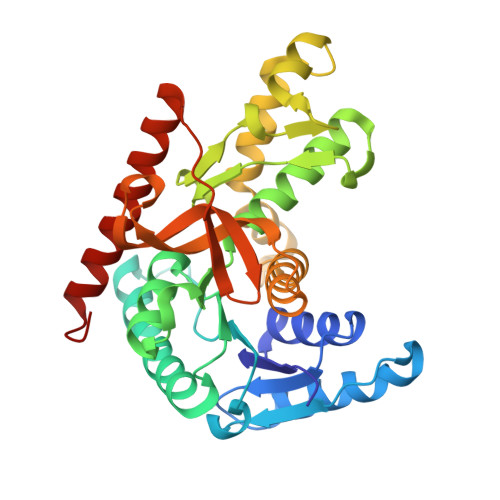
Conformational changes on substrate binding revealed by structures of Methylobacterium extorquens malate dehydrogenase.

Gonzalez, J.M.Marti-Arbona, R.Chen, J.C.H.Broom-Peltz, B.Unkefer, C.J.

(2018) Acta Crystallogr F Struct Biol Commun 74: 610-616

  • DOI: https://doi.org/10.1107/S2053230X18011809
  • Primary Citation Related Structures: 
    5UJK, 5ULV

  • PubMed Abstract: 

    Three high-resolution X-ray crystal structures of malate dehydrogenase (MDH; EC 1.1.1.37) from the methylotroph Methylobacterium extorquens AM1 are presented. By comparing the structures of apo MDH, a binary complex of MDH and NAD + , and a ternary complex of MDH and oxaloacetate with ADP-ribose occupying the pyridine nucleotide-binding site, conformational changes associated with the formation of the catalytic complex were characterized. While the substrate-binding site is accessible in the enzyme resting state or NAD + -bound forms, the substrate-bound form exhibits a closed conformation. This conformational change involves the transition of an α-helix to a 3 10 -helix, which causes the adjacent loop to close the active site following coenzyme and substrate binding. In the ternary complex, His284 forms a hydrogen bond to the C2 carbonyl of oxaloacetate, placing it in a position to donate a proton in the formation of (2S)-malate.


  • Organizational Affiliation
    • Instituto de Bionanotecnología del NOA, Consejo Nacional de Investigaciones Científicas y Técnicas, Universidad Nacional de Santiago del Estero, G4206XCP Santiago del Estero, Argentina.

Macromolecule Content 

  • Total Structure Weight: 33.76 kDa 
  • Atom Count: 2,776 
  • Modeled Residue Count: 318 
  • Deposited Residue Count: 320 
  • Unique protein chains: 1

Macromolecules

Find similar proteins by:|  3D Structure
Entity ID: 1
MoleculeChains  Sequence LengthOrganismDetailsImage
Malate dehydrogenase320Methylorubrum extorquensMutation(s): 0 
Gene Names: mdh
EC: 1.1.1.37
UniProt
Find proteins for A9W386 (Methylorubrum extorquens (strain PA1))
Explore A9W386 
Go to UniProtKB:  A9W386
Entity Groups
Sequence Clusters30% Identity50% Identity70% Identity90% Identity95% Identity100% Identity
UniProt GroupA9W386
Sequence Annotations
Expand
Reference Sequence

Experimental Data & Validation

Experimental Data

  • Method: X-RAY DIFFRACTION
  • Resolution: 1.66 Å
  • R-Value Free:  0.168 (Depositor), 0.168 (DCC) 
  • R-Value Work:  0.140 (Depositor), 0.139 (DCC) 
  • R-Value Observed: 0.141 (Depositor) 
Space Group: P 64 2 2
Unit Cell:
Length ( Å )Angle ( ˚ )
a = 108.995α = 90
b = 108.995β = 90
c = 104.659γ = 120
Software Package:
Software NamePurpose
REFMACrefinement
iMOSFLMdata reduction
Aimlessdata scaling
PHASERphasing

Structure Validation

View Full Validation Report


Entry History 

Deposition Data

  • Released Date: 2017-02-08 
  • Deposition Author(s): Gonzalez, J.M.
  • This entry supersedes: 4ROR

Revision History  (Full details and data files)

  • Version 1.0: 2017-02-08
    Type: Initial release
  • Version 1.1: 2017-08-23
    Changes: Advisory, Data collection
  • Version 1.2: 2018-11-14
    Changes: Data collection, Database references, Derived calculations
  • Version 1.3: 2018-11-21
    Changes: Data collection, Database references
  • Version 1.4: 2023-10-04
    Changes: Data collection, Database references, Derived calculations, Refinement description