5UJX

Crystal structure of DHFR in 20% Isopropanol


Experimental Data Snapshot

  • Method: X-RAY DIFFRACTION
  • Resolution: 1.80 Å
  • R-Value Free: 0.253 
  • R-Value Work: 0.207 
  • R-Value Observed: 0.209 

wwPDB Validation   3D Report Full Report


Ligand Structure Quality Assessment 


This is version 1.2 of the entry. See complete history

Re-refinement Note

This entry reflects an alternative modeling of the original data in: 5E8Q


Literature

Modulating Enzyme Activity by Altering Protein Dynamics with Solvent.

Duff Jr., M.R.Borreguero, J.M.Cuneo, M.J.Ramanathan, A.He, J.Kamath, G.Chennubhotla, S.C.Meilleur, F.Howell, E.E.Herwig, K.W.Myles, D.A.A.Agarwal, P.K.

(2018) Biochemistry 57: 4263-4275

  • DOI: https://doi.org/10.1021/acs.biochem.8b00424
  • Primary Citation of Related Structures:  
    5EAJ, 5UJX

  • PubMed Abstract: 

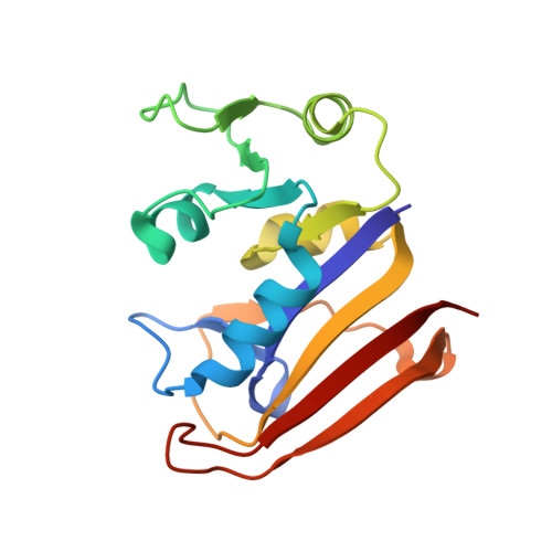
    Optimal enzyme activity depends on a number of factors, including structure and dynamics. The role of enzyme structure is well recognized; however, the linkage between protein dynamics and enzyme activity has given rise to a contentious debate. We have developed an approach that uses an aqueous mixture of organic solvent to control the functionally relevant enzyme dynamics (without changing the structure), which in turn modulates the enzyme activity. Using this approach, we predicted that the hydride transfer reaction catalyzed by the enzyme dihydrofolate reductase (DHFR) from Escherichia coli in aqueous mixtures of isopropanol (IPA) with water will decrease by ∼3 fold at 20% (v/v) IPA concentration. Stopped-flow kinetic measurements find that the pH-independent k hydride rate decreases by 2.2 fold. X-ray crystallographic enzyme structures show no noticeable differences, while computational studies indicate that the transition state and electrostatic effects were identical for water and mixed solvent conditions; quasi-elastic neutron scattering studies show that the dynamical enzyme motions are suppressed. Our approach provides a unique avenue to modulating enzyme activity through changes in enzyme dynamics. Further it provides vital insights that show the altered motions of DHFR cause significant changes in the enzyme's ability to access its functionally relevant conformational substates, explaining the decreased k hydride rate. This approach has important implications for obtaining fundamental insights into the role of rate-limiting dynamics in catalysis and as well as for enzyme engineering.


  • Organizational Affiliation

    Biochemistry & Cellular and Molecular Biology Department , University of Tennessee , Knoxville , Tennessee , United States.


Macromolecules
Find similar proteins by:  (by identity cutoff)  |  3D Structure
Entity ID: 1
MoleculeChains Sequence LengthOrganismDetailsImage
Dihydrofolate reductaseA [auth B],
B [auth A]
159Escherichia coliMutation(s): 1 
Gene Names: 
EC: 1.5.1.3
UniProt
Find proteins for P0ABQ4 (Escherichia coli (strain K12))
Explore P0ABQ4 
Go to UniProtKB:  P0ABQ4
Entity Groups  
Sequence Clusters30% Identity50% Identity70% Identity90% Identity95% Identity100% Identity
UniProt GroupP0ABQ4
Sequence Annotations
Expand
  • Reference Sequence
Small Molecules
Ligands 4 Unique
IDChains Name / Formula / InChI Key2D Diagram3D Interactions
FOL
Query on FOL

Download Ideal Coordinates CCD File 
C [auth B],
I [auth A]
FOLIC ACID
C19 H19 N7 O6
OVBPIULPVIDEAO-LBPRGKRZSA-N
IPA
Query on IPA

Download Ideal Coordinates CCD File 
D [auth B]ISOPROPYL ALCOHOL
C3 H8 O
KFZMGEQAYNKOFK-UHFFFAOYSA-N
CA
Query on CA

Download Ideal Coordinates CCD File 
E [auth B]CALCIUM ION
Ca
BHPQYMZQTOCNFJ-UHFFFAOYSA-N
CL
Query on CL

Download Ideal Coordinates CCD File 
F [auth B],
G [auth B],
H [auth B],
J [auth A]
CHLORIDE ION
Cl
VEXZGXHMUGYJMC-UHFFFAOYSA-M
Experimental Data & Validation

Experimental Data

  • Method: X-RAY DIFFRACTION
  • Resolution: 1.80 Å
  • R-Value Free: 0.253 
  • R-Value Work: 0.207 
  • R-Value Observed: 0.209 
  • Space Group: P 61
Unit Cell:
Length ( Å )Angle ( ˚ )
a = 91.946α = 90
b = 91.946β = 90
c = 73.088γ = 120
Software Package:
Software NamePurpose
HKL-2000data collection
HKL-2000data scaling
PHASERphasing
PHENIXrefinement
PDB_EXTRACTdata extraction
HKL-2000data reduction
HKL-2000data scaling
PHASERphasing
HKLdata reduction
HKLdata scaling

Structure Validation

View Full Validation Report


Ligand Structure Quality Assessment 


Entry History 

Deposition Data

Revision History  (Full details and data files)

  • Version 1.0: 2017-12-27
    Type: Initial release
  • Version 1.1: 2018-09-12
    Changes: Data collection, Database references
  • Version 1.2: 2023-10-04
    Changes: Data collection, Database references, Derived calculations, Refinement description