5UIW

Crystal Structure of CC Chemokine Receptor 5 (CCR5) in complex with high potency HIV entry inhibitor 5P7-CCL5


Experimental Data Snapshot

  • Method: X-RAY DIFFRACTION
  • Resolution: 2.20 Å
  • R-Value Free: 0.250 
  • R-Value Work: 0.216 
  • R-Value Observed: 0.218 

wwPDB Validation   3D Report Full Report


Ligand Structure Quality Assessment 


This is version 2.1 of the entry. See complete history


Literature

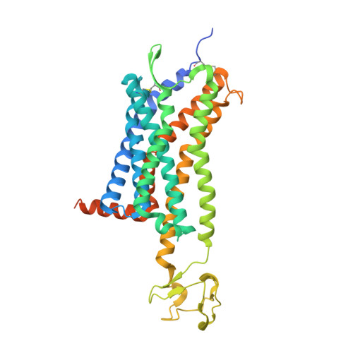
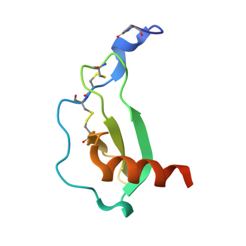
Structure of CC Chemokine Receptor 5 with a Potent Chemokine Antagonist Reveals Mechanisms of Chemokine Recognition and Molecular Mimicry by HIV.

Zheng, Y.Han, G.W.Abagyan, R.Wu, B.Stevens, R.C.Cherezov, V.Kufareva, I.Handel, T.M.

(2017) Immunity 46: 1005-1017.e5

  • DOI: https://doi.org/10.1016/j.immuni.2017.05.002
  • Primary Citation of Related Structures:  
    5UIW

  • PubMed Abstract: 

    CCR5 is the primary chemokine receptor utilized by HIV to infect leukocytes, whereas CCR5 ligands inhibit infection by blocking CCR5 engagement with HIV gp120. To guide the design of improved therapeutics, we solved the structure of CCR5 in complex with chemokine antagonist [5P7]CCL5. Several structural features appeared to contribute to the anti-HIV potency of [5P7]CCL5, including the distinct chemokine orientation relative to the receptor, the near-complete occupancy of the receptor binding pocket, the dense network of intermolecular hydrogen bonds, and the similarity of binding determinants with the FDA-approved HIV inhibitor Maraviroc. Molecular modeling indicated that HIV gp120 mimicked the chemokine interaction with CCR5, providing an explanation for the ability of CCR5 to recognize diverse ligands and gp120 variants. Our findings reveal that structural plasticity facilitates receptor-chemokine specificity and enables exploitation by HIV, and provide insight into the design of small molecule and protein inhibitors for HIV and other CCR5-mediated diseases.


  • Organizational Affiliation

    University of California, San Diego, Skaggs School of Pharmacy and Pharmaceutical Sciences, La Jolla, CA 92093, USA.


Macromolecules
Find similar proteins by:  (by identity cutoff)  |  3D Structure
Entity ID: 1
MoleculeChains Sequence LengthOrganismDetailsImage
C-C chemokine receptor type 5,Rubredoxin chimera411Homo sapiensClostridium pasteurianumMutation(s): 4 
Gene Names: CCR5CMKBR5
Membrane Entity: Yes 
UniProt & NIH Common Fund Data Resources
Find proteins for P51681 (Homo sapiens)
Explore P51681 
Go to UniProtKB:  P51681
PHAROS:  P51681
GTEx:  ENSG00000160791 
Find proteins for P00268 (Clostridium pasteurianum)
Explore P00268 
Go to UniProtKB:  P00268
Entity Groups  
Sequence Clusters30% Identity50% Identity70% Identity90% Identity95% Identity100% Identity
UniProt GroupsP00268P51681
Sequence Annotations
Expand
  • Reference Sequence
Find similar proteins by:  (by identity cutoff)  |  3D Structure
Entity ID: 2
MoleculeChains Sequence LengthOrganismDetailsImage
C-C motif chemokine 575Homo sapiensMutation(s): 0 
Gene Names: CCL5D17S136ESCYA5
Membrane Entity: Yes 
UniProt & NIH Common Fund Data Resources
Find proteins for P13501 (Homo sapiens)
Explore P13501 
Go to UniProtKB:  P13501
PHAROS:  P13501
GTEx:  ENSG00000271503 
Entity Groups  
Sequence Clusters30% Identity50% Identity70% Identity90% Identity95% Identity100% Identity
UniProt GroupP13501
Sequence Annotations
Expand
  • Reference Sequence
Small Molecules
Ligands 3 Unique
IDChains Name / Formula / InChI Key2D Diagram3D Interactions
OLC
Query on OLC

Download Ideal Coordinates CCD File 
D [auth A]
E [auth A]
F [auth A]
G [auth A]
H [auth A]
D [auth A],
E [auth A],
F [auth A],
G [auth A],
H [auth A],
I [auth A]
(2R)-2,3-dihydroxypropyl (9Z)-octadec-9-enoate
C21 H40 O4
RZRNAYUHWVFMIP-GDCKJWNLSA-N
OLA
Query on OLA

Download Ideal Coordinates CCD File 
J [auth A],
K [auth A]
OLEIC ACID
C18 H34 O2
ZQPPMHVWECSIRJ-KTKRTIGZSA-N
ZN
Query on ZN

Download Ideal Coordinates CCD File 
C [auth A]ZINC ION
Zn
PTFCDOFLOPIGGS-UHFFFAOYSA-N
Modified Residues  1 Unique
IDChains TypeFormula2D DiagramParent
PCA
Query on PCA
B
L-PEPTIDE LINKINGC5 H7 N O3GLN
Experimental Data & Validation

Experimental Data

  • Method: X-RAY DIFFRACTION
  • Resolution: 2.20 Å
  • R-Value Free: 0.250 
  • R-Value Work: 0.216 
  • R-Value Observed: 0.218 
  • Space Group: P 2 21 21
Unit Cell:
Length ( Å )Angle ( ˚ )
a = 38.01α = 90
b = 52.64β = 90
c = 302.67γ = 90
Software Package:
Software NamePurpose
PHENIXrefinement
Aimlessdata scaling
PHASERphasing
PDB_EXTRACTdata extraction
XDSdata reduction

Structure Validation

View Full Validation Report


Ligand Structure Quality Assessment 


Entry History & Funding Information

Deposition Data


Funding OrganizationLocationGrant Number
National Institutes of Health/National Institute Of Allergy and Infectious Diseases (NIH/NIAID)United StatesAI118985

Revision History  (Full details and data files)

  • Version 1.0: 2017-06-28
    Type: Initial release
  • Version 1.1: 2017-09-13
    Changes: Author supporting evidence
  • Version 1.2: 2018-04-18
    Changes: Data collection, Database references
  • Version 2.0: 2019-12-11
    Changes: Author supporting evidence, Polymer sequence
  • Version 2.1: 2023-10-04
    Changes: Data collection, Database references, Derived calculations, Refinement description