5UIO

structure of DHFR with bound DAP, p-ABG and NADP


Experimental Data Snapshot

  • Method: X-RAY DIFFRACTION
  • Resolution: 1.93 Å
  • R-Value Free: 0.230 
  • R-Value Work: 0.192 
  • R-Value Observed: 0.193 

wwPDB Validation   3D Report Full Report


Ligand Structure Quality Assessment 


This is version 1.1 of the entry. See complete history


Literature

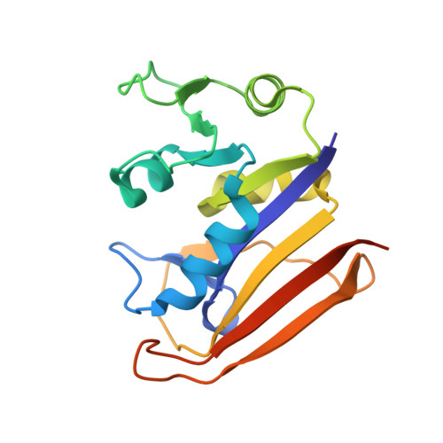
A Structural Basis for Biguanide Activity.

Gabel, S.A.Duff, M.R.Pedersen, L.C.DeRose, E.F.Krahn, J.M.Howell, E.E.London, R.E.

(2017) Biochemistry 56: 4786-4798

  • DOI: https://doi.org/10.1021/acs.biochem.7b00619
  • Primary Citation of Related Structures:  
    5UIH, 5UII, 5UIO, 5UIP

  • PubMed Abstract: 

    Metformin is the most commonly prescribed treatment for type II diabetes and related disorders; however, molecular insights into its mode(s) of action have been limited by an absence of structural data. Structural considerations along with a growing body of literature demonstrating its effects on one-carbon metabolism suggest the possibility of folate mimicry and anti-folate activity. Motivated by the growing recognition that anti-diabetic biguanides may act directly upon the gut microbiome, we have determined structures of the complexes formed between the anti-diabetic biguanides (phenformin, buformin, and metformin) and Escherichia coli dihydrofolate reductase (ecDHFR) based on nuclear magnetic resonance, crystallographic, and molecular modeling studies. Interligand Overhauser effects indicate that metformin can form ternary complexes with p-aminobenzoyl-l-glutamate (pABG) as well as other ligands that occupy the region of the folate-binding site that interacts with pABG; however, DHFR inhibition is not cooperative. The biguanides competitively inhibit the activity of ecDHFR, with the phenformin inhibition constant being 100-fold lower than that of metformin. This inhibition may be significant at concentrations present in the gut of treated individuals, and inhibition of DHFR in intestinal mucosal cells may also occur if accumulation levels are sufficient. Perturbation of folate homeostasis can alter the pyridine nucleotide redox ratios that are important regulators of cellular metabolism.


  • Organizational Affiliation

    Genome Integrity and Structural Biology Laboratory, National Institute of Environmental Health Sciences, National Institutes of Health , 111 T. W. Alexander Drive, Research Triangle Park, North Carolina 27709, United States.


Macromolecules
Find similar proteins by:  (by identity cutoff)  |  3D Structure
Entity ID: 1
MoleculeChains Sequence LengthOrganismDetailsImage
Dihydrofolate reductase
A, B, C, D, E
170Escherichia coliMutation(s): 0 
Gene Names: folA
EC: 1.5.1.3
UniProt
Find proteins for P0ABQ4 (Escherichia coli (strain K12))
Explore P0ABQ4 
Go to UniProtKB:  P0ABQ4
Entity Groups  
Sequence Clusters30% Identity50% Identity70% Identity90% Identity95% Identity100% Identity
UniProt GroupP0ABQ4
Sequence Annotations
Expand
  • Reference Sequence
Small Molecules
Ligands 5 Unique
IDChains Name / Formula / InChI Key2D Diagram3D Interactions
NAP
Query on NAP

Download Ideal Coordinates CCD File 
H [auth A],
N [auth C],
S [auth D],
W [auth E]
NADP NICOTINAMIDE-ADENINE-DINUCLEOTIDE PHOSPHATE
C21 H28 N7 O17 P3
XJLXINKUBYWONI-NNYOXOHSSA-N
8DM
Query on 8DM

Download Ideal Coordinates CCD File 
G [auth A],
M [auth C],
R [auth D],
V [auth E]
N-(4-aminobenzene-1-carbonyl)-L-glutamic acid
C12 H14 N2 O5
GADGMZDHLQLZRI-VIFPVBQESA-N
LG3
Query on LG3

Download Ideal Coordinates CCD File 
F [auth A],
I [auth B],
L [auth C],
Q [auth D],
U [auth E]
PYRIMIDINE-2,4-DIAMINE
C4 H6 N4
YAAWASYJIRZXSZ-UHFFFAOYSA-N
BME
Query on BME

Download Ideal Coordinates CCD File 
J [auth B],
O [auth C],
T [auth D],
X [auth E]
BETA-MERCAPTOETHANOL
C2 H6 O S
DGVVWUTYPXICAM-UHFFFAOYSA-N
FMT
Query on FMT

Download Ideal Coordinates CCD File 
K [auth B],
P [auth C]
FORMIC ACID
C H2 O2
BDAGIHXWWSANSR-UHFFFAOYSA-N
Experimental Data & Validation

Experimental Data

  • Method: X-RAY DIFFRACTION
  • Resolution: 1.93 Å
  • R-Value Free: 0.230 
  • R-Value Work: 0.192 
  • R-Value Observed: 0.193 
  • Space Group: P 21 21 21
Unit Cell:
Length ( Å )Angle ( ˚ )
a = 62.781α = 90
b = 103.945β = 90
c = 145.4γ = 90
Software Package:
Software NamePurpose
PHENIXrefinement
HKL-3000data reduction
HKL-3000data scaling
PHENIXphasing

Structure Validation

View Full Validation Report


Ligand Structure Quality Assessment 


Entry History 

Deposition Data

Revision History  (Full details and data files)

  • Version 1.0: 2017-11-22
    Type: Initial release
  • Version 1.1: 2023-10-04
    Changes: Data collection, Database references, Refinement description