5UA3

Crystal Structure of a DNA G-Quadruplex with a Cytosine Bulge


Experimental Data Snapshot

  • Method: X-RAY DIFFRACTION
  • Resolution: 1.88 Å
  • R-Value Free: 0.247 
  • R-Value Work: 0.227 
  • R-Value Observed: 0.229 

wwPDB Validation   3D Report Full Report


This is version 1.6 of the entry. See complete history


Literature

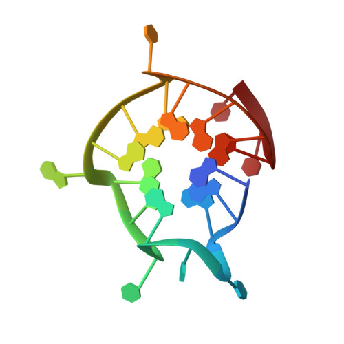
Structure and hydrodynamics of a DNA G-quadruplex with a cytosine bulge.

Meier, M.Moya-Torres, A.Krahn, N.J.McDougall, M.D.Orriss, G.L.McRae, E.K.S.Booy, E.P.McEleney, K.Patel, T.R.McKenna, S.A.Stetefeld, J.

(2018) Nucleic Acids Res 46: 5319-5331

  • DOI: https://doi.org/10.1093/nar/gky307
  • Primary Citation of Related Structures:  
    5UA3

  • PubMed Abstract: 

    The identification of four-stranded G-quadruplexes (G4s) has highlighted the fact that DNA has additional spatial organisations at its disposal other than double-stranded helices. Recently, it became clear that the formation of G4s is not limited to the traditional G3+NL1G3+NL2G3+NL3G3+ sequence motif. Instead, the G3 triplets can be interrupted by deoxythymidylate (DNA) or uridylate (RNA) where the base forms a bulge that loops out from the G-quadruplex core. Here, we report the first high-resolution X-ray structure of a unique unimolecular DNA G4 with a cytosine bulge. The G4 forms a dimer that is stacked via its 5'-tetrads. Analytical ultracentrifugation, static light scattering and small angle X-ray scattering confirmed that the G4 adapts a predominantly dimeric structure in solution. We provide a comprehensive comparison of previously published G4 structures containing bulges and report a special γ torsion angle range preferentially populated by the G4 core guanylates adjacent to bulges. Since the penalty for introducing bulges appears to be negligible, it should be possible to functionalize G4s by introducing artificial or modified nucleotides at such positions. The presence of the bulge alters the surface of the DNA, providing an opportunity to develop drugs that can specifically target individual G4s.


  • Organizational Affiliation

    Department of Chemistry, University of Manitoba, Winnipeg, Manitoba R3T 2N2, Canada.


Macromolecules

Find similar nucleic acids by:  Sequence   |   3D Structure  

Entity ID: 1
MoleculeChains LengthOrganismImage
DNA (5'-D(*GP*GP*GP*TP*TP*GP*CP*GP*GP*AP*GP*GP*GP*TP*GP*GP*GP*CP*CP*T)-3')
A, B
20Homo sapiens
Sequence Annotations
Expand
  • Reference Sequence
Experimental Data & Validation

Experimental Data

  • Method: X-RAY DIFFRACTION
  • Resolution: 1.88 Å
  • R-Value Free: 0.247 
  • R-Value Work: 0.227 
  • R-Value Observed: 0.229 
  • Space Group: P 1
Unit Cell:
Length ( Å )Angle ( ˚ )
a = 30.103α = 64.65
b = 33.1β = 78.64
c = 33.282γ = 81.78
Software Package:
Software NamePurpose
REFMACrefinement
DENZOdata reduction
SCALEPACKdata scaling
PHASERphasing
PDB_EXTRACTdata extraction
PHASERphasing

Structure Validation

View Full Validation Report


Entry History & Funding Information

Deposition Data


Funding OrganizationLocationGrant Number
Natural Sciences and Engineering Research Council (NSERC, Canada)Canada342077-2012
Natural Sciences and Engineering Research Council (NSERC, Canada)Canada345517-07
Cancer Research SocietyCanada--

Revision History  (Full details and data files)

  • Version 1.0: 2017-11-22
    Type: Initial release
  • Version 1.1: 2017-12-06
    Changes: Database references
  • Version 1.2: 2018-05-09
    Changes: Data collection, Database references, Structure summary
  • Version 1.3: 2018-05-16
    Changes: Data collection, Database references
  • Version 1.4: 2018-06-13
    Changes: Data collection, Database references
  • Version 1.5: 2020-01-08
    Changes: Author supporting evidence
  • Version 1.6: 2023-10-04
    Changes: Data collection, Database references, Derived calculations, Refinement description