


Cocrystal structure of the intermembrane space region of the plastid division proteins PARC6 and PDV1
Delmar, J.A., Yu, E.W., Osteryoung, K.W.To be published.
Experimental Data Snapshot
wwPDB Validation 3D Report Full Report

Entity ID: 1 | |||||
|---|---|---|---|---|---|
| Molecule | Chains | Sequence Length | Organism | Details | Image |
| PARALOG OF ACCUMULATION AND REPLICATION OF CHLOROPLASTS 6 (PARC6) | 233 | Arabidopsis thaliana | Mutation(s): 0 Gene Names: CDP1, ARC6H, PARC6, At3g19180, MVI11.9 | 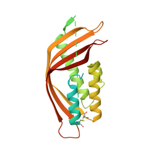 | |
UniProt | |||||
Entity Groups | |||||
| Sequence Clusters | 30% Identity50% Identity70% Identity90% Identity95% Identity100% Identity | ||||
| UniProt Group | Q8VY16 | ||||
Sequence AnnotationsExpand | |||||
Reference Sequence | |||||
| Ligands 1 Unique | |||||
|---|---|---|---|---|---|
| ID | Chains | Name / Formula / InChI Key | 2D Diagram | 3D Interactions | |
| MG Download:Ideal Coordinates CCD File | C [auth A], D [auth B] | MAGNESIUM ION Mg JLVVSXFLKOJNIY-UHFFFAOYSA-N |  | ||
| Length ( Å ) | Angle ( ˚ ) |
|---|---|
| a = 44.533 | α = 90 |
| b = 124.546 | β = 90 |
| c = 130.046 | γ = 90 |
| Software Name | Purpose |
|---|---|
| SCALEPACK | data scaling |
| PHENIX | refinement |
| PDB_EXTRACT | data extraction |
| Coot | model building |
| MOLREP | phasing |


