5U91 | pdb_00005u91

Crystal structure of Tre/loxLTR complex


Experimental Data Snapshot

  • Method: X-RAY DIFFRACTION
  • Resolution: 3.10 Å
  • R-Value Free: 
    0.253 (Depositor), 0.243 (DCC) 
  • R-Value Work: 
    0.209 (Depositor) 
  • R-Value Observed: 
    0.211 (Depositor) 

Starting Model: experimental
View more details

wwPDB Validation 3D Report Full Report

Validation slider image for 5U91

This is version 1.3 of the entry. See complete history

Literature

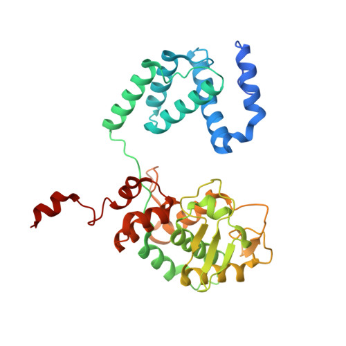
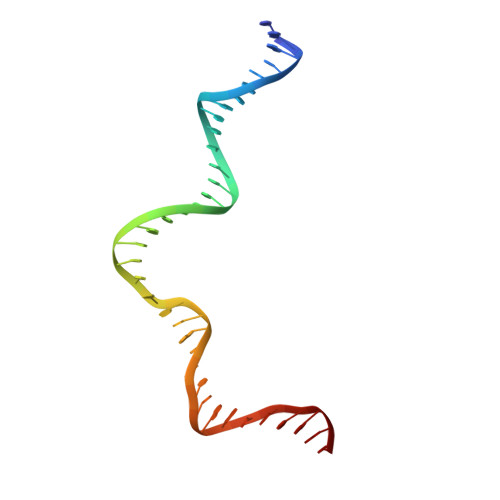
Crystal structure of an engineered, HIV-specific recombinase for removal of integrated proviral DNA.

Meinke, G.Karpinski, J.Buchholz, F.Bohm, A.

(2017) Nucleic Acids Res 45: 9726-9740

  • DOI: https://doi.org/10.1093/nar/gkx603
  • Primary Citation Related Structures: 
    5U91

  • PubMed Abstract: 

    As part of the HIV infection cycle, viral DNA inserts into the genome of host cells such that the integrated DNA encoding the viral proteins is flanked by long terminal repeat (LTR) regions from the retrovirus. In an effort to develop novel genome editing techniques that safely excise HIV provirus from cells, Tre, an engineered version of Cre recombinase, was designed to target a 34-bp sequence within the HIV-1 LTR (loxLTR). The sequence targeted by Tre lacks the symmetry present in loxP, the natural DNA substrate for Cre. We report here the crystal structure of a catalytically inactive (Y324F) mutant of this engineered Tre recombinase in complex with the loxLTR DNA substrate. We also report that 17 of the 19 amino acid changes relative to Cre contribute to the altered specificity, even though many of these residues do not contact the DNA directly. We hypothesize that some mutations increase the flexibility of the Cre tetramer and that this, along with flexibility in the DNA, enable the engineered enzyme and DNA substrate to adopt complementary conformations.


  • Organizational Affiliation
    • Department of Developmental, Molecular and Chemical Biology, Tufts University School of Medicine, Boston, MA 02111, USA.

Macromolecule Content 

  • Total Structure Weight: 200.71 kDa 
  • Atom Count: 13,259 
  • Modeled Residue Count: 1,451 
  • Deposited Residue Count: 1,528 
  • Unique protein chains: 1
  • Unique nucleic acid chains: 2

Macromolecules


Find similar proteins by:|  3D Structure
Entity ID: 1
MoleculeChains  Sequence LengthOrganismDetailsImage
Tre recombinase proteinA,
B,
C [auth E],
D [auth F]
345synthetic constructMutation(s): 0 
Find similar nucleic acids by:  (by identity cutoff) 
Entity ID: 2
MoleculeChains LengthOrganismImage
DNA (37-MER)E [auth C],
G
37Human immunodeficiency virus 1
Sequence Annotations
Expand
Reference Sequence
Find similar nucleic acids by:  (by identity cutoff) 
Entity ID: 3
MoleculeChains LengthOrganismImage
DNA (37-MER)F [auth D],
H
37Human immunodeficiency virus 1
Sequence Annotations
Expand
Reference Sequence

Experimental Data & Validation

Experimental Data

  • Method: X-RAY DIFFRACTION
  • Resolution: 3.10 Å
  • R-Value Free:  0.253 (Depositor), 0.243 (DCC) 
  • R-Value Work:  0.209 (Depositor) 
  • R-Value Observed: 0.211 (Depositor) 
Space Group: P 1 21 1
Unit Cell:
Length ( Å )Angle ( ˚ )
a = 70.26α = 90
b = 193.39β = 111.25
c = 89.02γ = 90
Software Package:
Software NamePurpose
HKL-2000data collection
SCALEPACKdata scaling
PHASERphasing
PHENIXrefinement
PDB_EXTRACTdata extraction
DENZOdata reduction

Structure Validation

View Full Validation Report


Entry History 

Deposition Data

Revision History  (Full details and data files)

  • Version 1.0: 2017-07-26
    Type: Initial release
  • Version 1.1: 2017-10-04
    Changes: Database references, Refinement description
  • Version 1.2: 2023-10-04
    Changes: Data collection, Database references, Refinement description
  • Version 1.3: 2024-11-06
    Changes: Structure summary