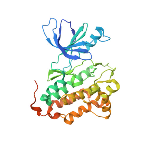
5U8L

Crystal structure of EGFR kinase domain in complex with a sulfonyl fluoride probe XO44


Experimental Data Snapshot

  • Method: X-RAY DIFFRACTION
  • Resolution: 1.60 Å
  • R-Value Free: 0.202 
  • R-Value Work: 0.189 
  • R-Value Observed: 0.190 

wwPDB Validation   3D Report Full Report


Ligand Structure Quality Assessment 


This is version 1.1 of the entry. See complete history


Literature

Broad-Spectrum Kinase Profiling in Live Cells with Lysine-Targeted Sulfonyl Fluoride Probes.

Zhao, Q.Ouyang, X.Wan, X.Gajiwala, K.S.Kath, J.C.Jones, L.H.Burlingame, A.L.Taunton, J.

(2017) J Am Chem Soc 139: 680-685

  • DOI: https://doi.org/10.1021/jacs.6b08536
  • Primary Citation of Related Structures:  
    5K9I, 5U8L

  • PubMed Abstract: 

    Protein kinases comprise a large family of structurally related enzymes. A major goal in kinase-inhibitor development is to selectively engage the desired kinase while avoiding myriad off-target kinases. However, quantifying inhibitor interactions with multiple endogenous kinases in live cells remains an unmet challenge. Here, we report the design of sulfonyl fluoride probes that covalently label a broad swath of the intracellular kinome with high efficiency. Protein crystallography and mass spectrometry confirmed a chemoselective reaction between the sulfonyl fluoride and a conserved lysine in the ATP binding site. Optimized probe 2 (XO44) covalently modified up to 133 endogenous kinases, efficiently competing with high intracellular concentrations of ATP. We employed probe 2 and label-free mass spectrometry to quantify intracellular kinase engagement by the approved drug, dasatinib. The data revealed saturable dasatinib binding to a small subset of kinase targets at clinically relevant concentrations, highlighting the utility of lysine-targeted sulfonyl fluoride probes in demanding chemoproteomic applications.


  • Organizational Affiliation

    Worldwide Research and Development, Pfizer , San Diego, California 92121, United States.


Macromolecules
Find similar proteins by:  (by identity cutoff)  |  3D Structure
Entity ID: 1
MoleculeChains Sequence LengthOrganismDetailsImage
Epidermal growth factor receptor329Homo sapiensMutation(s): 0 
Gene Names: EGFRERBBERBB1HER1
EC: 2.7.10.1
UniProt & NIH Common Fund Data Resources
Find proteins for P00533 (Homo sapiens)
Explore P00533 
Go to UniProtKB:  P00533
PHAROS:  P00533
GTEx:  ENSG00000146648 
Entity Groups  
Sequence Clusters30% Identity50% Identity70% Identity90% Identity95% Identity100% Identity
UniProt GroupP00533
Sequence Annotations
Expand
  • Reference Sequence
Small Molecules
Ligands 3 Unique
IDChains Name / Formula / InChI Key2D Diagram3D Interactions
O44
Query on O44

Download Ideal Coordinates CCD File 
B [auth A]4-[(4-{4-[(3-cyclopropyl-1H-pyrazol-5-yl)amino]-6-[(prop-2-yn-1-yl)carbamoyl]pyrimidin-2-yl}piperazin-1-yl)methyl]benzene-1-sulfonyl fluoride
C25 H27 F N8 O3 S
ZBSPMOBILDLOCV-UHFFFAOYSA-N
SO4
Query on SO4

Download Ideal Coordinates CCD File 
C [auth A]SULFATE ION
O4 S
QAOWNCQODCNURD-UHFFFAOYSA-L
GOL
Query on GOL

Download Ideal Coordinates CCD File 
D [auth A]GLYCEROL
C3 H8 O3
PEDCQBHIVMGVHV-UHFFFAOYSA-N
Experimental Data & Validation

Experimental Data

  • Method: X-RAY DIFFRACTION
  • Resolution: 1.60 Å
  • R-Value Free: 0.202 
  • R-Value Work: 0.189 
  • R-Value Observed: 0.190 
  • Space Group: P 21 21 21
Unit Cell:
Length ( Å )Angle ( ˚ )
a = 40.364α = 90
b = 69.977β = 90
c = 110.403γ = 90
Software Package:
Software NamePurpose
CNXrefinement
XDSdata reduction
Aimlessdata scaling
CNXphasing

Structure Validation

View Full Validation Report


Ligand Structure Quality Assessment 


Entry History 

Deposition Data

Revision History  (Full details and data files)

  • Version 1.0: 2017-01-25
    Type: Initial release
  • Version 1.1: 2017-02-01
    Changes: Database references