5U7V

Crystal structure of a nucleoside triphosphate diphosphohydrolase (NTPDase) from the legume Trifolium repens in complex with AMP


Experimental Data Snapshot

  • Method: X-RAY DIFFRACTION
  • Resolution: 2.15 Å
  • R-Value Free: 0.265 
  • R-Value Work: 0.195 
  • R-Value Observed: 0.199 

wwPDB Validation   3D Report Full Report


Ligand Structure Quality Assessment 


This is version 1.4 of the entry. See complete history


Literature

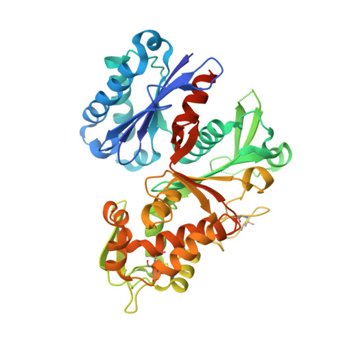
Structures and kinetics for plant nucleoside triphosphate diphosphohydrolases support a domain motion catalytic mechanism.

Summers, E.L.Cumming, M.H.Oulavallickal, T.Roberts, N.J.Arcus, V.L.

(2017) Protein Sci 26: 1627-1638

  • DOI: https://doi.org/10.1002/pro.3199
  • Primary Citation of Related Structures:  
    5U7P, 5U7V, 5U7W, 5U7X

  • PubMed Abstract: 

    Extracellular nucleoside triphosphate diphosphohydrolases (NTPDases) are enzymes that hydrolyze extracellular nucleotides to the respective monophosphate nucleotides. In the past 20 years, NTPDases belonging to mammalian, parasitic and prokaryotic domains of life have been discovered, cloned and characterized. We reveal the first structures of NTPDases from the legume plant species Trifolium repens (7WC) and Vigna unguiculata subsp. cylindrica (DbLNP). Four crystal structures of 7WC and DbLNP were determined at resolutions between 1.9 and 2.6 Å. For 7WC, structures were determined for an -apo form (1.89 Å) and with the product AMP (2.15 Å) and adenine and phosphate (1.76 Å) bound. For DbLNP, a structure was solved with phosphate and manganese bound (2.60 Å). Thorough kinetic data and analysis is presented. The structure of 7WC and DbLNP reveals that these NTPDases can adopt two conformations depending on the molecule and co-factor bound in the active site. A central hinge region creates a "butterfly-like" motion of the domains that reduces the width of the inter-domain active site cleft upon molecule binding. This phenomenon has been previously described in Rattus norvegicus and Legionella pneumophila NTPDaseI and Toxoplasma gondii NTPDaseIII suggesting a common catalytic mechanism across the domains of life.


  • Organizational Affiliation

    School of Science, University of Waikato, Private Bag 3105, Hamilton, 3240, New Zealand.


Macromolecules
Find similar proteins by:  (by identity cutoff)  |  3D Structure
Entity ID: 1
MoleculeChains Sequence LengthOrganismDetailsImage
Apyrase426Trifolium repensMutation(s): 0 
EC: 3.6.1.5
UniProt
Find proteins for B9U139 (Trifolium repens)
Explore B9U139 
Go to UniProtKB:  B9U139
Entity Groups  
Sequence Clusters30% Identity50% Identity70% Identity90% Identity95% Identity100% Identity
UniProt GroupB9U139
Sequence Annotations
Expand
  • Reference Sequence
Small Molecules
Ligands 1 Unique
IDChains Name / Formula / InChI Key2D Diagram3D Interactions
AMP
Query on AMP

Download Ideal Coordinates CCD File 
B [auth A]ADENOSINE MONOPHOSPHATE
C10 H14 N5 O7 P
UDMBCSSLTHHNCD-KQYNXXCUSA-N
Experimental Data & Validation

Experimental Data

  • Method: X-RAY DIFFRACTION
  • Resolution: 2.15 Å
  • R-Value Free: 0.265 
  • R-Value Work: 0.195 
  • R-Value Observed: 0.199 
  • Space Group: P 1 21 1
Unit Cell:
Length ( Å )Angle ( ˚ )
a = 53.83α = 90
b = 51.91β = 108.46
c = 69.46γ = 90
Software Package:
Software NamePurpose
REFMACrefinement
MOSFLMdata reduction
SCALAdata scaling
MOLREPphasing

Structure Validation

View Full Validation Report


Ligand Structure Quality Assessment 


Entry History 

Deposition Data

Revision History  (Full details and data files)

  • Version 1.0: 2017-05-31
    Type: Initial release
  • Version 1.1: 2017-06-07
    Changes: Database references
  • Version 1.2: 2017-06-14
    Changes: Database references
  • Version 1.3: 2017-08-02
    Changes: Database references
  • Version 1.4: 2023-10-04
    Changes: Data collection, Database references, Refinement description