


Crystal Structure of the holo Domain-Swapped Dimer mutant Q108K:K40D Human Cellular Retinol Binding Protein II bound with all trans retinal
Assar, Z., Geiger, J.H.To be published.
Experimental Data Snapshot
Starting Model: experimental
View more details
Macromolecule Content
Entity ID: 1 | |||||
|---|---|---|---|---|---|
| Molecule | Chains | Sequence Length | Organism | Details | Image |
| Retinol-binding protein 2 | 133 | Homo sapiens | Mutation(s): 2 Gene Names: RBP2, CRBP2 | 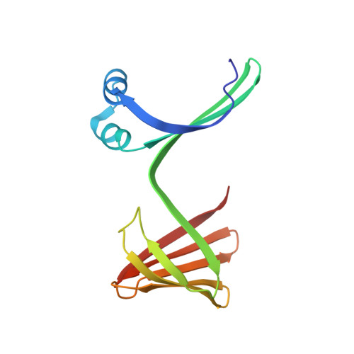 | |
UniProt & NIH Common Fund Data Resources | |||||
PHAROS: P50120 GTEx: ENSG00000114113 | |||||
Entity Groups | |||||
| Sequence Clusters | 30% Identity50% Identity70% Identity90% Identity95% Identity100% Identity | ||||
| UniProt Group | P50120 | ||||
Sequence AnnotationsExpand | |||||
Reference Sequence | |||||
Entity ID: 2 | |||||
|---|---|---|---|---|---|
| Molecule | Chains | Sequence Length | Organism | Details | Image |
| Retinol-binding protein 2 | H [auth G], I | 133 | Homo sapiens | Mutation(s): 1 Gene Names: RBP2, CRBP2 | 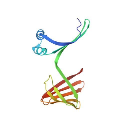 |
UniProt & NIH Common Fund Data Resources | |||||
PHAROS: P50120 GTEx: ENSG00000114113 | |||||
Entity Groups | |||||
| Sequence Clusters | 30% Identity50% Identity70% Identity90% Identity95% Identity100% Identity | ||||
| UniProt Group | P50120 | ||||
Sequence AnnotationsExpand | |||||
Reference Sequence | |||||
| Ligands 1 Unique | |||||
|---|---|---|---|---|---|
| ID | Chains | Name / Formula / InChI Key | 2D Diagram | 3D Interactions | |
| RET Download:Ideal Coordinates CCD File | M [auth A] N [auth B] O [auth C] P [auth D] Q [auth H] | RETINAL C20 H28 O NCYCYZXNIZJOKI-OVSJKPMPSA-N |  | ||
| Length ( Å ) | Angle ( ˚ ) |
|---|---|
| a = 65.474 | α = 90 |
| b = 73.597 | β = 90 |
| c = 351.601 | γ = 90 |
| Software Name | Purpose |
|---|---|
| PHENIX | refinement |
| PHENIX | phasing |
| HKL-2000 | data scaling |
| PHENIX | model building |
| HKL-2000 | data reduction |
| Funding Organization | Location | Grant Number |
|---|---|---|
| National Institutes of Health/National Institute of General Medical Sciences (NIH/NIGMS) | United States | -- |


