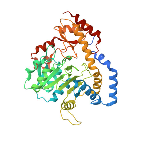
5U24

X-ray structure of the WlaRG aminotransferase from campylobacter jejuni, K184A mutant in complex with TDP-Fuc3N


Experimental Data Snapshot

  • Method: X-RAY DIFFRACTION
  • Resolution: 1.70 Å
  • R-Value Free: 0.146 
  • R-Value Work: 0.105 
  • R-Value Observed: 0.107 

wwPDB Validation   3D Report Full Report


Ligand Structure Quality Assessment 


This is version 1.4 of the entry. See complete history


Literature

Structural investigation on WlaRG from Campylobacter jejuni: A sugar aminotransferase.

Dow, G.T.Gilbert, M.Thoden, J.B.Holden, H.M.

(2017) Protein Sci 26: 586-599

  • DOI: https://doi.org/10.1002/pro.3109
  • Primary Citation of Related Structures:  
    3NYS, 3NYT, 3NYU, 5U1Z, 5U20, 5U21, 5U23, 5U24

  • PubMed Abstract: 

    Campylobacter jejuni is a Gram-negative bacterium that represents a leading cause of human gastroenteritis worldwide. Of particular concern is the link between C. jejuni infections and the subsequent development of Guillain-Barré syndrome, an acquired autoimmune disorder leading to paralysis. All Gram-negative bacteria contain complex glycoconjugates anchored to their outer membranes, but in most strains of C. jejuni, this lipoglycan lacks the O-antigen repeating units. Recent mass spectrometry analyses indicate that the C. jejuni 81116 (Penner serotype HS:6) lipoglycan contains two dideoxyhexosamine residues, and enzymological assay data show that this bacterial strain can synthesize both dTDP-3-acetamido-3,6-dideoxy-d-glucose and dTDP-3-acetamido-3,6-dideoxy-d-galactose. The focus of this investigation is on WlaRG from C. jejuni, which plays a key role in the production of these unusual sugars by functioning as a pyridoxal 5'-phosphate dependent aminotransferase. Here, we describe the first three-dimensional structures of the enzyme in various complexes determined to resolutions of 1.7 Å or higher. Of particular significance are the external aldimine structures of WlaRG solved in the presence of either dTDP-3-amino-3,6-dideoxy-d-galactose or dTDP-3-amino-3,6-dideoxy-d-glucose. These models highlight the manner in which WlaRG can accommodate sugars with differing stereochemistries about their C-4' carbon positions. In addition, we present a corrected structure of WbpE, a related sugar aminotransferase from Pseudomonas aeruginosa, solved to 1.3 Å resolution.


  • Organizational Affiliation

    Department of Biochemistry, University of Wisconsin, Madison, Wisconsin, 53706.


Macromolecules
Find similar proteins by:  (by identity cutoff)  |  3D Structure
Entity ID: 1
MoleculeChains Sequence LengthOrganismDetailsImage
Putative aminotransferase
A, B, C, D
382Campylobacter jejuniMutation(s): 1 
UniProt
Find proteins for Q9ALS9 (Campylobacter jejuni)
Explore Q9ALS9 
Go to UniProtKB:  Q9ALS9
Entity Groups  
Sequence Clusters30% Identity50% Identity70% Identity90% Identity95% Identity100% Identity
UniProt GroupQ9ALS9
Sequence Annotations
Expand
  • Reference Sequence
Small Molecules
Ligands 4 Unique
IDChains Name / Formula / InChI Key2D Diagram3D Interactions
7SG
Query on 7SG

Download Ideal Coordinates CCD File 
F [auth A],
K [auth B],
O [auth C],
Q [auth D]
(2R,3R,4S,5R,6R)-3,5-dihydroxy-4-[(E)-({3-hydroxy-2-methyl-5-[(phosphonooxy)methyl]pyridin-4-yl}methylidene)amino]-6-methyltetrahydro-2H-pyran-2-yl [(2R,3S,5R)-3-hydroxy-5-(5-methyl-2,4-dioxo-3,4-dihydropyrimidin-1(2H)-yl)tetrahydrofuran-2-yl]methyl dihydrogen diphosphate (non-preferred name)
C24 H35 N4 O19 P3
UJDWKFAFGYHSFM-QBLVGKJYSA-N
EDO
Query on EDO

Download Ideal Coordinates CCD File 
G [auth A]
H [auth A]
I [auth A]
L [auth B]
M [auth B]
G [auth A],
H [auth A],
I [auth A],
L [auth B],
M [auth B],
P [auth C],
R [auth D],
S [auth D]
1,2-ETHANEDIOL
C2 H6 O2
LYCAIKOWRPUZTN-UHFFFAOYSA-N
CL
Query on CL

Download Ideal Coordinates CCD File 
J [auth A]CHLORIDE ION
Cl
VEXZGXHMUGYJMC-UHFFFAOYSA-M
NA
Query on NA

Download Ideal Coordinates CCD File 
E [auth A],
N [auth C]
SODIUM ION
Na
FKNQFGJONOIPTF-UHFFFAOYSA-N
Experimental Data & Validation

Experimental Data

  • Method: X-RAY DIFFRACTION
  • Resolution: 1.70 Å
  • R-Value Free: 0.146 
  • R-Value Work: 0.105 
  • R-Value Observed: 0.107 
  • Space Group: P 1 21 1
Unit Cell:
Length ( Å )Angle ( ˚ )
a = 109.057α = 90
b = 56.795β = 90.02
c = 124.306γ = 90
Software Package:
Software NamePurpose
REFMACrefinement
SAINTdata reduction
SADABSdata scaling
PHASERphasing

Structure Validation

View Full Validation Report


Ligand Structure Quality Assessment 


Entry History & Funding Information

Deposition Data


Funding OrganizationLocationGrant Number
National Institutes of Health/National Institute of General Medical Sciences (NIH/NIGMS)United StatesGM115921

Revision History  (Full details and data files)

  • Version 1.0: 2017-01-11
    Type: Initial release
  • Version 1.1: 2017-03-08
    Changes: Database references
  • Version 1.2: 2017-09-27
    Changes: Author supporting evidence
  • Version 1.3: 2020-01-01
    Changes: Author supporting evidence
  • Version 1.4: 2023-10-04
    Changes: Data collection, Database references, Derived calculations, Refinement description