5TVM

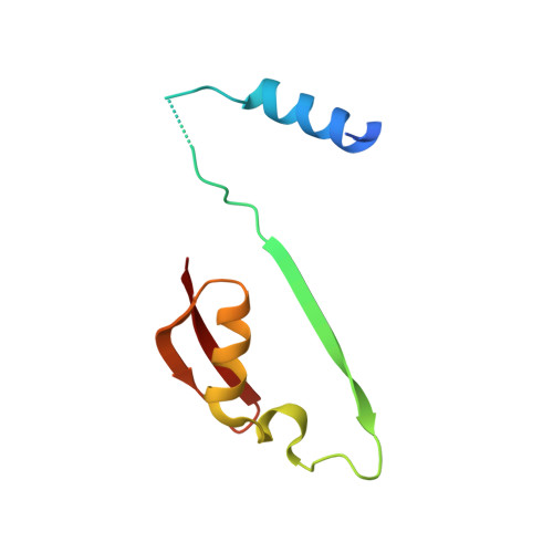
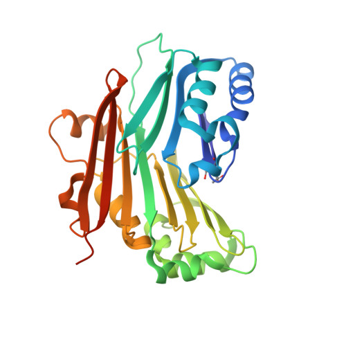
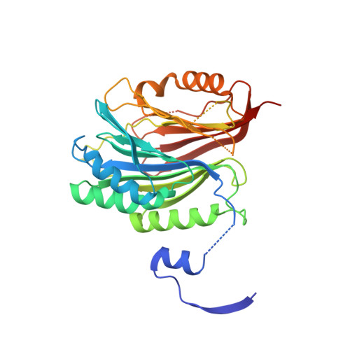
Crystal structure of Trypanosoma brucei AdoMetDC/prozyme heterodimer


Experimental Data Snapshot

  • Method: X-RAY DIFFRACTION
  • Resolution: 2.41 Å
  • R-Value Free: 0.274 
  • R-Value Work: 0.229 
  • R-Value Observed: 0.231 

wwPDB Validation   3D Report Full Report


This is version 2.0 of the entry. See complete history


Literature

Relief of autoinhibition by conformational switch explains enzyme activation by a catalytically dead paralog.

Volkov, O.A.Kinch, L.N.Ariagno, C.Deng, X.Zhong, S.Grishin, N.V.Tomchick, D.R.Chen, Z.Phillips, M.A.

(2016) Elife 5

  • DOI: https://doi.org/10.7554/eLife.20198
  • Primary Citation of Related Structures:  
    5TVF, 5TVM, 5TVO

  • PubMed Abstract: 

    Catalytically inactive enzyme paralogs occur in many genomes. Some regulate their active counterparts but the structural principles of this regulation remain largely unknown. We report X-ray structures of Trypanosoma brucei S -adenosylmethionine decarboxylase alone and in functional complex with its catalytically dead paralogous partner, prozyme. We show monomeric Tb AdoMetDC is inactive because of autoinhibition by its N-terminal sequence. Heterodimerization with prozyme displaces this sequence from the active site through a complex mechanism involving a cis -to- trans proline isomerization, reorganization of a β-sheet, and insertion of the N-terminal α-helix into the heterodimer interface, leading to enzyme activation. We propose that the evolution of this intricate regulatory mechanism was facilitated by the acquisition of the dimerization domain, a single step that can in principle account for the divergence of regulatory schemes in the AdoMetDC enzyme family. These studies elucidate an allosteric mechanism in an enzyme and a plausible scheme by which such complex cooperativity evolved.


  • Organizational Affiliation

    Department of Pharmacology, University of Texas Southwestern Medical Center, Dallas, United States.


Macromolecules
Find similar proteins by:  (by identity cutoff)  |  3D Structure
Entity ID: 1
MoleculeChains Sequence LengthOrganismDetailsImage
S-adenosylmethionine decarboxylase beta chain
A, C
85Trypanosoma brucei brucei TREU927Mutation(s): 0 
Gene Names: Tb927.6.4410Tb927.6.4460
EC: 4.1.1.50
UniProt
Find proteins for Q587A7 (Trypanosoma brucei brucei (strain 927/4 GUTat10.1))
Explore Q587A7 
Go to UniProtKB:  Q587A7
Entity Groups  
Sequence Clusters30% Identity50% Identity70% Identity90% Identity95% Identity100% Identity
UniProt GroupQ587A7
Sequence Annotations
Expand
  • Reference Sequence
Find similar proteins by:  (by identity cutoff)  |  3D Structure
Entity ID: 2
MoleculeChains Sequence LengthOrganismDetailsImage
S-adenosylmethionine decarboxylase alpha chain
B, D
285Trypanosoma brucei brucei TREU927Mutation(s): 0 
Gene Names: Tb927.6.4410Tb927.6.4460
EC: 4.1.1.50
UniProt
Find proteins for Q587A7 (Trypanosoma brucei brucei (strain 927/4 GUTat10.1))
Explore Q587A7 
Go to UniProtKB:  Q587A7
Entity Groups  
Sequence Clusters30% Identity50% Identity70% Identity90% Identity95% Identity100% Identity
UniProt GroupQ587A7
Sequence Annotations
Expand
  • Reference Sequence
Find similar proteins by:  (by identity cutoff)  |  3D Structure
Entity ID: 3
MoleculeChains Sequence LengthOrganismDetailsImage
S-adenosylmethionine decarboxylase proenzyme-like, putative
E, F
325Trypanosoma brucei brucei TREU927Mutation(s): 0 
Gene Names: Tb927.6.4470
EC: 4.1.1.50
UniProt
Find proteins for Q587B3 (Trypanosoma brucei brucei (strain 927/4 GUTat10.1))
Explore Q587B3 
Go to UniProtKB:  Q587B3
Entity Groups  
Sequence Clusters30% Identity50% Identity70% Identity90% Identity95% Identity100% Identity
UniProt GroupQ587B3
Sequence Annotations
Expand
  • Reference Sequence
Experimental Data & Validation

Experimental Data

  • Method: X-RAY DIFFRACTION
  • Resolution: 2.41 Å
  • R-Value Free: 0.274 
  • R-Value Work: 0.229 
  • R-Value Observed: 0.231 
  • Space Group: P 1 21 1
Unit Cell:
Length ( Å )Angle ( ˚ )
a = 81.297α = 90
b = 96.708β = 102.64
c = 99.584γ = 90
Software Package:
Software NamePurpose
PHENIXrefinement
HKL-3000data reduction
HKL-3000data scaling
PHASERphasing

Structure Validation

View Full Validation Report


Entry History & Funding Information

Deposition Data


Funding OrganizationLocationGrant Number
National Institutes of Health/National Institute Of Allergy and Infectious Diseases (NIH/NIAID)United States2R37AI034432
National Institutes of Health/National Institute Of Allergy and Infectious Diseases (NIH/NIAID)United StatesR01AI090599

Revision History  (Full details and data files)

  • Version 1.0: 2017-01-11
    Type: Initial release
  • Version 1.1: 2017-09-27
    Changes: Advisory, Author supporting evidence
  • Version 1.2: 2019-12-11
    Changes: Advisory, Author supporting evidence
  • Version 1.3: 2023-10-04
    Changes: Data collection, Database references, Derived calculations, Refinement description
  • Version 2.0: 2023-11-15
    Changes: Atomic model, Data collection, Derived calculations