5TTR | pdb_00005ttr

LEU 55 PRO TRANSTHYRETIN CRYSTAL STRUCTURE


Experimental Data Snapshot

  • Method: X-RAY DIFFRACTION
  • Resolution: 2.70 Å
  • R-Value Work: 
    0.199 (Depositor) 

Starting Model: experimental
View more details

wwPDB Validation 3D Report Full Report

Validation slider image for 5TTR

This is version 1.5 of the entry. See complete history

Literature

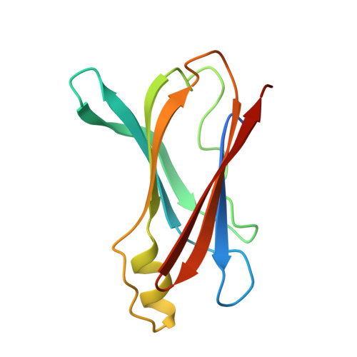
The crystal structure of amyloidogenic Leu55 --> Pro transthyretin variant reveals a possible pathway for transthyretin polymerization into amyloid fibrils.

Sebastiao, M.P.Saraiva, M.J.Damas, A.M.

(1998) J Biological Chem 273: 24715-24722

  • DOI: https://doi.org/10.1074/jbc.273.38.24715
  • Primary Citation Related Structures: 
    5TTR

  • PubMed Abstract: 

    The x-ray crystal structure of the amyloidogenic Leu55 --> Pro transthyretin (TTR) variant, implicated as the causative agent in early-onset familial amyloidotic polyneuropathy (Jacobson, D. R., McFarlin, D. E., Kane, I., and Buxbaum, J. N. (1992) Hum. Genet. 89, 353-356), has been solved by molecular replacement, refined at 2.7 A to a Rcryst value of 0.190 (Fobs > 2.0sigma), and compared with wild-type transthyretin to understand the molecular mechanism(s) involved in amyloidogenesis. Leu55 --> Pro TTR crystallizes in space group C2, with eight monomers in the asymmetric unit, and the observed packing contacts are considerably different from those described for the wild-type protein. Refinement of the crystal structure shows that the proline for leucine substitution disrupts the hydrogen bonds between strands D and A, resulting in different interface contacts. Based on the assumption that the observed packing contacts may be significant for amyloidogenesis, a model for the TTR amyloid is proposed. It consists of a tubular structure with inner and outer diameters approximately of 30 and 100 A and four monomers per cross-section.


  • Organizational Affiliation
    • Instituto de Biologia Molecular e Celular, Rua do Campo Alegre, no. 823, 4150 Porto, Portugal.

Macromolecule Content 

  • Total Structure Weight: 110.09 kDa 
  • Atom Count: 7,147 
  • Modeled Residue Count: 928 
  • Deposited Residue Count: 1,016 
  • Unique protein chains: 1

Macromolecules

Find similar proteins by:|  3D Structure
Entity ID: 1
MoleculeChains  Sequence LengthOrganismDetailsImage
TRANSTHYRETIN
A, B, C, D, E
A, B, C, D, E, F, G, H
127Homo sapiensMutation(s): 1 
UniProt & NIH Common Fund Data Resources
Find proteins for P02766 (Homo sapiens)
Explore P02766 
Go to UniProtKB:  P02766
PHAROS:  P02766
GTEx:  ENSG00000118271 
Entity Groups
Sequence Clusters30% Identity50% Identity70% Identity90% Identity95% Identity100% Identity
UniProt GroupP02766
Sequence Annotations
Expand
Reference Sequence

Experimental Data & Validation

Experimental Data

  • Method: X-RAY DIFFRACTION
  • Resolution: 2.70 Å
  • R-Value Work:  0.199 (Depositor) 
Space Group: C 1 2 1
Unit Cell:
Length ( Å )Angle ( ˚ )
a = 149.99α = 90
b = 78.74β = 100.5
c = 98.95γ = 90
Software Package:
Software NamePurpose
AMoREphasing
PROLSQrefinement
DENZOdata reduction
SCALEPACKdata scaling

Structure Validation

View Full Validation Report


Entry History 

Deposition Data

Revision History  (Full details and data files)

  • Version 1.0: 1999-06-01
    Type: Initial release
  • Version 1.1: 2008-03-25
    Changes: Version format compliance
  • Version 1.2: 2011-07-13
    Changes: Version format compliance
  • Version 1.3: 2021-11-03
    Changes: Data collection, Database references
  • Version 1.4: 2023-08-09
    Changes: Refinement description
  • Version 1.5: 2024-05-22
    Changes: Data collection