5TLC | pdb_00005tlc

Crystal structure of BdsA from Bacillus subtilis WU-S2B


Experimental Data Snapshot

  • Method: X-RAY DIFFRACTION
  • Resolution: 2.80 Å
  • R-Value Free: 
    0.274 (Depositor), 0.273 (DCC) 
  • R-Value Work: 
    0.212 (Depositor), 0.214 (DCC) 
  • R-Value Observed: 
    0.215 (Depositor) 

wwPDB Validation 3D Report Full Report

Validation slider image for 5TLC

This is version 1.2 of the entry. See complete history

Literature

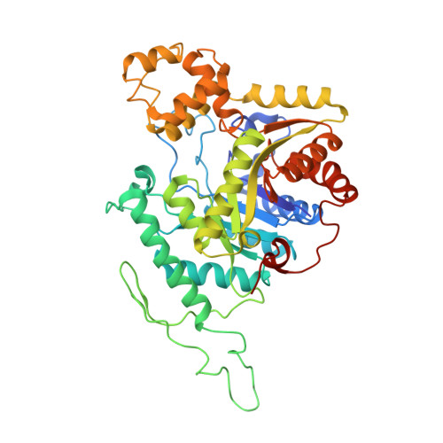
Crystal structure of dibenzothiophene sulfone monooxygenase BdsA from Bacillus subtilis WU-S2B

Okai, M.Lee, W.C.Guan, L.J.Ohshiro, T.Izumi, Y.Tanokura, M.

(2017) Proteins 85: 1171-1177

  • DOI: https://doi.org/10.1002/prot.25267
  • Primary Citation Related Structures: 
    5TLC

  • PubMed Abstract: 

    The dibenzothiophene (DBT) sulfone monooxygenase BdsA from Bacillus subtilis WU-S2B catalyzes the conversion of DBT sulfone to 2'-hydroxybiphenyl 2-sulfinate. We report the crystal structures of BdsA at a resolution of 2.80 Å. BdsA exists as a homotetramer with a dimer-of-dimers configuration in the crystal, and the interaction between E288 and R296 in BdsA is important for tetramer formation. A structural comparison with homologous proteins shows that the orientation and location of the α9-α12 helices in BdsA are closer to those of the closed form than those of the open form in the EDTA monooxygenase EmoA. Proteins 2017; 85:1171-1177. © 2017 Wiley Periodicals, Inc.


  • Organizational Affiliation
    • Department of Applied Biological Chemistry, Graduate School of Agricultural and Life Sciences, University of Tokyo, Bunkyo-ku, Tokyo, 113-8657, Japan.

Macromolecule Content 

  • Total Structure Weight: 198.84 kDa 
  • Atom Count: 12,884 
  • Modeled Residue Count: 1,657 
  • Deposited Residue Count: 1,812 
  • Unique protein chains: 1

Macromolecules

Find similar proteins by:|  3D Structure
Entity ID: 1
MoleculeChains  Sequence LengthOrganismDetailsImage
Dibenzothiophene desulfurization enzyme A
A, B, C, D
453Bacillus subtilisMutation(s): 0 
Gene Names: bdsA
UniProt
Find proteins for Q8GRC7 (Bacillus subtilis)
Explore Q8GRC7 
Go to UniProtKB:  Q8GRC7
Entity Groups
Sequence Clusters30% Identity50% Identity70% Identity90% Identity95% Identity100% Identity
UniProt GroupQ8GRC7
Sequence Annotations
Expand
Reference Sequence

Experimental Data & Validation

Experimental Data

  • Method: X-RAY DIFFRACTION
  • Resolution: 2.80 Å
  • R-Value Free:  0.274 (Depositor), 0.273 (DCC) 
  • R-Value Work:  0.212 (Depositor), 0.214 (DCC) 
  • R-Value Observed: 0.215 (Depositor) 
Space Group: P 32
Unit Cell:
Length ( Å )Angle ( ˚ )
a = 84.126α = 90
b = 84.126β = 90
c = 269.456γ = 120
Software Package:
Software NamePurpose
REFMACrefinement
XDSdata reduction
SCALAdata scaling

Structure Validation

View Full Validation Report


Entry History 

Deposition Data

Revision History  (Full details and data files)

  • Version 1.0: 2017-05-03
    Type: Initial release
  • Version 1.1: 2017-05-31
    Changes: Database references
  • Version 1.2: 2024-03-20
    Changes: Data collection, Database references