5TK5

Crystal structure of human 3HAO with iron bound in the active site


Experimental Data Snapshot

  • Method: X-RAY DIFFRACTION
  • Resolution: 1.88 Å
  • R-Value Free: 0.218 
  • R-Value Work: 0.203 
  • R-Value Observed: 0.203 

wwPDB Validation   3D Report Full Report


This is version 1.4 of the entry. See complete history


Literature

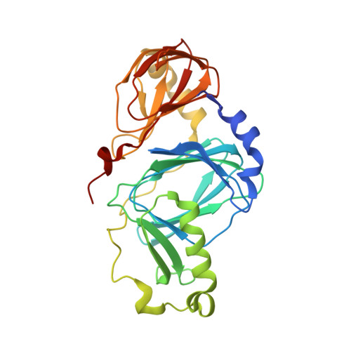
Crystal structures of human 3-hydroxyanthranilate 3,4-dioxygenase with native and non-native metals bound in the active site.

Pidugu, L.S.Neu, H.Wong, T.L.Pozharski, E.Molloy, J.L.Michel, S.L.Toth, E.A.

(2017) Acta Crystallogr D Struct Biol 73: 340-348

  • DOI: https://doi.org/10.1107/S2059798317002029
  • Primary Citation of Related Structures:  
    5TK5, 5TKQ

  • PubMed Abstract: 

    3-Hydroxyanthranilate 3,4-dioxygenase (3HAO) is an enzyme in the microglial branch of the kynurenine pathway of tryptophan degradation. 3HAO is a non-heme iron-containing, ring-cleaving extradiol dioxygenase that catalyzes the addition of both atoms of O 2 to the kynurenine pathway metabolite 3-hydroxyanthranilic acid (3-HANA) to form quinolinic acid (QUIN). QUIN is a highly potent excitotoxin that has been implicated in a number of neurodegenerative conditions, making 3HAO a target for pharmacological downregulation. Here, the first crystal structure of human 3HAO with the native iron bound in its active site is presented, together with an additional structure with zinc (a known inhibitor of human 3HAO) bound in the active site. The metal-binding environment is examined both structurally and via inductively coupled plasma mass spectrometry (ICP-MS), X-ray fluorescence spectroscopy (XRF) and electron paramagnetic resonance spectroscopy (EPR). The studies identified Met35 as the source of potential new interactions with substrates and inhibitors, which may prove useful in future therapeutic efforts.


  • Organizational Affiliation

    Department of Biochemistry and Molecular Biology, University of Maryland School of Medicine, Baltimore, MD 21201, USA.


Macromolecules
Find similar proteins by:  (by identity cutoff)  |  3D Structure
Entity ID: 1
MoleculeChains Sequence LengthOrganismDetailsImage
3-hydroxyanthranilate 3,4-dioxygenase290Homo sapiensMutation(s): 0 
Gene Names: HAAO
EC: 1.13.11.6
UniProt & NIH Common Fund Data Resources
Find proteins for P46952 (Homo sapiens)
Explore P46952 
Go to UniProtKB:  P46952
PHAROS:  P46952
GTEx:  ENSG00000162882 
Entity Groups  
Sequence Clusters30% Identity50% Identity70% Identity90% Identity95% Identity100% Identity
UniProt GroupP46952
Sequence Annotations
Expand
  • Reference Sequence
Experimental Data & Validation

Experimental Data

  • Method: X-RAY DIFFRACTION
  • Resolution: 1.88 Å
  • R-Value Free: 0.218 
  • R-Value Work: 0.203 
  • R-Value Observed: 0.203 
  • Space Group: P 65 2 2
Unit Cell:
Length ( Å )Angle ( ˚ )
a = 57.076α = 90
b = 57.076β = 90
c = 415.871γ = 120
Software Package:
Software NamePurpose
BUSTERrefinement
XDSdata reduction
Aimlessdata scaling
PHASERphasing

Structure Validation

View Full Validation Report


Entry History & Funding Information

Deposition Data


Funding OrganizationLocationGrant Number
National Science Foundation (NSF, United States)United StatesCHE1306208

Revision History  (Full details and data files)

  • Version 1.0: 2017-04-12
    Type: Initial release
  • Version 1.1: 2017-09-27
    Changes: Author supporting evidence
  • Version 1.2: 2018-04-18
    Changes: Data collection, Database references
  • Version 1.3: 2019-11-27
    Changes: Author supporting evidence, Derived calculations
  • Version 1.4: 2023-10-04
    Changes: Data collection, Database references, Refinement description