5TJX | pdb_00005tjx

Structure of human plasma kallikrein


Experimental Data Snapshot

  • Method: X-RAY DIFFRACTION
  • Resolution: 1.41 Å
  • R-Value Free: 
    0.184 (Depositor) 
  • R-Value Work: 
    0.163 (Depositor) 
  • R-Value Observed: 
    0.163 (Depositor) 

Starting Model: experimental
View more details

wwPDB Validation 3D Report Full Report

Validation slider image for 5TJX

Ligand Structure Quality Assessment 


This is version 1.3 of the entry. See complete history

Literature

Structure-Guided Design of Novel, Potent, and Selective Macrocyclic Plasma Kallikrein Inhibitors.

Li, Z.Partridge, J.Silva-Garcia, A.Rademacher, P.Betz, A.Xu, Q.Sham, H.Hu, Y.Shan, Y.Liu, B.Zhang, Y.Shi, H.Xu, Q.Ma, X.Zhang, L.

(2017) ACS Med Chem Lett 8: 185-190

  • DOI: https://doi.org/10.1021/acsmedchemlett.6b00384
  • Primary Citation Related Structures: 
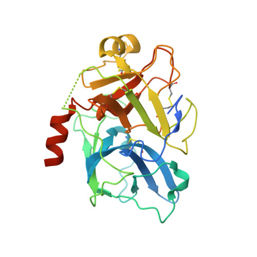
    5TJX

  • PubMed Abstract: 

    A series of macrocyclic analogues were designed and synthesized based on the cocrystal structure of small molecule plasma kallikrein (pKal) inhibitor, 2 , with the pKal protease domain. This led to the discovery of a potent macrocyclic pKal inhibitor 29 , with an IC 50 of 2 nM for one olefinic isomer and 42.3 nM for the other olefinic isomer.


  • Organizational Affiliation
    • Global Blood Therapeutics , South San Francisco, California 94080, United States.

Macromolecule Content 

  • Total Structure Weight: 30.14 kDa 
  • Atom Count: 2,244 
  • Modeled Residue Count: 231 
  • Deposited Residue Count: 263 
  • Unique protein chains: 1

Macromolecules

Find similar proteins by:|  3D Structure
Entity ID: 1
MoleculeChains  Sequence LengthOrganismDetailsImage
Plasma kallikrein263Homo sapiensMutation(s): 4 
Gene Names: KLKB1KLK3
EC: 3.4.21.34
UniProt & NIH Common Fund Data Resources
Find proteins for P03952 (Homo sapiens)
Explore P03952 
Go to UniProtKB:  P03952
PHAROS:  P03952
GTEx:  ENSG00000164344 
Entity Groups
Sequence Clusters30% Identity50% Identity70% Identity90% Identity95% Identity100% Identity
UniProt GroupP03952
Sequence Annotations
Expand
Reference Sequence

Small Molecules

Ligands 2 Unique
IDChains Name / Formula / InChI Key2D Diagram3D Interactions
GBT

Query on GBT


Download:Ideal Coordinates CCD File
B [auth A](8E)-3-amino-1-methyl-15-[(1H-pyrazol-1-yl)methyl]-7,10,11,12,24,25-hexahydro-6H,18H,23H-19,22-(metheno)pyrido[4,3-j][1,9,13,17,18]benzodioxatriazacyclohenicosin-23-one
C29 H33 N7 O3
ZVYLYYBRLJOHMS-DUXPYHPUSA-N
PO4

Query on PO4


Download:Ideal Coordinates CCD File
C [auth A],
D [auth A]
PHOSPHATE ION
O4 P
NBIIXXVUZAFLBC-UHFFFAOYSA-K
Binding Affinity Annotations 
IDSourceBinding Affinity
GBT BindingDB:  5TJX IC50: 2 (nM) from 1 assay(s)

Experimental Data & Validation

Experimental Data

  • Method: X-RAY DIFFRACTION
  • Resolution: 1.41 Å
  • R-Value Free:  0.184 (Depositor) 
  • R-Value Work:  0.163 (Depositor) 
  • R-Value Observed: 0.163 (Depositor) 
Space Group: P 21 21 21
Unit Cell:
Length ( Å )Angle ( ˚ )
a = 55.66α = 90
b = 59.62β = 90
c = 77.11γ = 90
Software Package:
Software NamePurpose
PHENIXrefinement
XDSdata reduction
SCALAdata scaling
PHASERphasing

Structure Validation

View Full Validation Report


Ligand Structure Quality Assessment 


Entry History 

Deposition Data

Revision History  (Full details and data files)

  • Version 1.0: 2016-12-21
    Type: Initial release
  • Version 1.1: 2017-03-01
    Changes: Database references
  • Version 1.2: 2023-10-04
    Changes: Data collection, Database references, Refinement description
  • Version 1.3: 2024-10-23
    Changes: Structure summary