5TBT

Crystal Structure of Isoform 2 of Purine Nucleoside Phosphorylase complexed with Cytidine


Experimental Data Snapshot

  • Method: X-RAY DIFFRACTION
  • Resolution: 2.10 Å
  • R-Value Free: 0.226 
  • R-Value Work: 0.190 
  • R-Value Observed: 0.192 

wwPDB Validation   3D Report Full Report


Ligand Structure Quality Assessment 


This is version 1.4 of the entry. See complete history


Literature

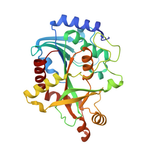
The molecular structure of Schistosoma mansoni PNP isoform 2 provides insights into the nucleoside selectivity of PNPs.

Torini, J.R.Romanello, L.Batista, F.A.H.Serrao, V.H.B.Faheem, M.Zeraik, A.E.Bird, L.Nettleship, J.Reddivari, Y.Owens, R.DeMarco, R.Borges, J.C.Brandao-Neto, J.Pereira, H.D.

(2018) PLoS One 13: e0203532-e0203532

  • DOI: https://doi.org/10.1371/journal.pone.0203532
  • Primary Citation of Related Structures:  
    5CXQ, 5CXS, 5KO5, 5TBS, 5TBT, 5TBU

  • PubMed Abstract: 

    Purine nucleoside phosphorylases (PNPs) play an important role in the blood fluke parasite Schistosoma mansoni as a key enzyme of the purine salvage pathway. Here we present the structural and kinetic characterization of a new PNP isoform from S. mansoni, SmPNP2. Thermofluorescence screening of different ligands suggested cytidine and cytosine are potential ligands. The binding of cytosine and cytidine were confirmed by isothermal titration calorimetry, with a KD of 27 μM for cytosine, and a KM of 76.3 μM for cytidine. SmPNP2 also displays catalytic activity against inosine and adenosine, making it the first described PNP with robust catalytic activity towards both pyrimidines and purines. Crystal structures of SmPNP2 with different ligands were obtained and comparison of these structures with the previously described S. mansoni PNP (SmPNP1) provided clues for the unique capacity of SmPNP2 to bind pyrimidines. When compared with the structure of SmPNP1, substitutions in the vicinity of SmPNP2 active site alter the architecture of the nucleoside base binding site thus permitting an alternative binding mode for nucleosides, with a 180° rotation from the canonical binding mode. The remarkable plasticity of this binding site enhances our understanding of the correlation between structure and nucleotide selectivity, thus suggesting new ways to analyse PNP activity.


  • Organizational Affiliation

    Laboratório de Biologia Estrutural, Instituto de Física de São Carlos, Universidade de São Paulo, São Carlos, São Paulo, Brazil.


Macromolecules
Find similar proteins by:  (by identity cutoff)  |  3D Structure
Entity ID: 1
MoleculeChains Sequence LengthOrganismDetailsImage
Purine nucleoside phosphorylase287Schistosoma mansoniMutation(s): 0 
EC: 2.4.2.1
UniProt
Find proteins for A0A0U3AGT1 (Schistosoma mansoni)
Explore A0A0U3AGT1 
Go to UniProtKB:  A0A0U3AGT1
Entity Groups  
Sequence Clusters30% Identity50% Identity70% Identity90% Identity95% Identity100% Identity
UniProt GroupA0A0U3AGT1
Sequence Annotations
Expand
  • Reference Sequence
Small Molecules
Ligands 2 Unique
IDChains Name / Formula / InChI Key2D Diagram3D Interactions
CTN
Query on CTN

Download Ideal Coordinates CCD File 
B [auth A]4-AMINO-1-BETA-D-RIBOFURANOSYL-2(1H)-PYRIMIDINONE
C9 H13 N3 O5
UHDGCWIWMRVCDJ-XVFCMESISA-N
SO4
Query on SO4

Download Ideal Coordinates CCD File 
C [auth A]SULFATE ION
O4 S
QAOWNCQODCNURD-UHFFFAOYSA-L
Experimental Data & Validation

Experimental Data

  • Method: X-RAY DIFFRACTION
  • Resolution: 2.10 Å
  • R-Value Free: 0.226 
  • R-Value Work: 0.190 
  • R-Value Observed: 0.192 
  • Space Group: P 21 3
Unit Cell:
Length ( Å )Angle ( ˚ )
a = 100.153α = 90
b = 100.153β = 90
c = 100.153γ = 90
Software Package:
Software NamePurpose
xia2data scaling
xia2data reduction
PHASERphasing
PHENIXrefinement
PDB_EXTRACTdata extraction

Structure Validation

View Full Validation Report


Ligand Structure Quality Assessment 


Entry History & Funding Information

Deposition Data


Funding OrganizationLocationGrant Number
Sao Paulo Research Foundation (FAPESP)Brazil2012/14223-9
Brazilian National Council for Scientific and Technological Development (CNPq)Brazil474402/2013-4

Revision History  (Full details and data files)

  • Version 1.0: 2017-10-11
    Type: Initial release
  • Version 1.1: 2018-09-26
    Changes: Data collection, Database references
  • Version 1.2: 2019-04-17
    Changes: Author supporting evidence, Data collection
  • Version 1.3: 2020-01-01
    Changes: Author supporting evidence
  • Version 1.4: 2023-10-04
    Changes: Data collection, Database references, Refinement description