


Crystal Structure of a S-adenosylmethionine Synthase from Neisseria gonorrhoeae with bound S-adenosylmethionine, AMP, Pyrophosphate, Phosphate, and Magnesium
Dranow, D.M., Delker, S.L., Lorimer, D.D., Edwards, T.E.To be published.
Experimental Data Snapshot
Starting Model: experimental
View more details
Entity ID: 1 | |||||
|---|---|---|---|---|---|
| Molecule | Chains | Sequence Length | Organism | Details | Image |
| S-adenosylmethionine synthase | 397 | Neisseria gonorrhoeae FA 1090 | Mutation(s): 0 Gene Names: metK, NGO0106 EC: 2.5.1.6 | 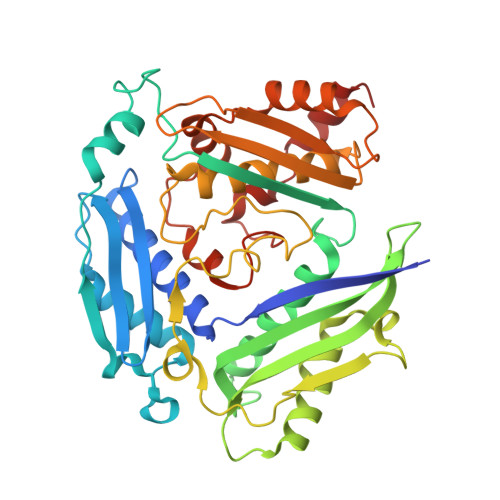 | |
UniProt | |||||
Entity Groups | |||||
| Sequence Clusters | 30% Identity50% Identity70% Identity90% Identity95% Identity100% Identity | ||||
| UniProt Group | Q5FAC0 | ||||
Sequence AnnotationsExpand | |||||
Reference Sequence | |||||
| Ligands 6 Unique | |||||
|---|---|---|---|---|---|
| ID | Chains | Name / Formula / InChI Key | 2D Diagram | 3D Interactions | |
| SAM Download:Ideal Coordinates CCD File | I [auth B] | S-ADENOSYLMETHIONINE C15 H22 N6 O5 S MEFKEPWMEQBLKI-FCKMPRQPSA-N |  | ||
| AMP Download:Ideal Coordinates CCD File | D [auth A] | ADENOSINE MONOPHOSPHATE C10 H14 N5 O7 P UDMBCSSLTHHNCD-KQYNXXCUSA-N |  | ||
| 3PO Download:Ideal Coordinates CCD File | K [auth B] | TRIPHOSPHATE H5 O10 P3 UNXRWKVEANCORM-UHFFFAOYSA-N |  | ||
| POP Download:Ideal Coordinates CCD File | F [auth A] | PYROPHOSPHATE 2- H2 O7 P2 XPPKVPWEQAFLFU-UHFFFAOYSA-L |  | ||
| PO4 Download:Ideal Coordinates CCD File | G [auth A] | PHOSPHATE ION O4 P NBIIXXVUZAFLBC-UHFFFAOYSA-K |  | ||
| MG Download:Ideal Coordinates CCD File | C [auth A], E [auth A], H [auth B], J [auth B] | MAGNESIUM ION Mg JLVVSXFLKOJNIY-UHFFFAOYSA-N |  | ||
| Length ( Å ) | Angle ( ˚ ) |
|---|---|
| a = 115.71 | α = 90 |
| b = 115.71 | β = 90 |
| c = 146.24 | γ = 90 |
| Software Name | Purpose |
|---|---|
| XSCALE | data scaling |
| PHENIX | refinement |
| PDB_EXTRACT | data extraction |
| XDS | data reduction |
| MR-Rosetta | phasing |


