5T5G | pdb_00005t5g

human SETD8 in complex with MS2177


Experimental Data Snapshot

  • Method: X-RAY DIFFRACTION
  • Resolution: 2.10 Å
  • R-Value Free: 
    0.227 (Depositor), 0.237 (DCC) 
  • R-Value Work: 
    0.205 (Depositor), 0.214 (DCC) 
  • R-Value Observed: 
    0.206 (Depositor) 

Starting Model: experimental
View more details

wwPDB Validation 3D Report Full Report

Validation slider image for 5T5G

Ligand Structure Quality Assessment 


This is version 1.5 of the entry. See complete history

Literature

Structure-Based Design of a Covalent Inhibitor of the SET Domain-Containing Protein 8 (SETD8) Lysine Methyltransferase.

Butler, K.V.Ma, A.Yu, W.Li, F.Tempel, W.Babault, N.Pittella-Silva, F.Shao, J.Wang, J.Luo, M.Vedadi, M.Brown, P.J.Arrowsmith, C.H.Jin, J.

(2016) J Med Chem 59: 9881-9889

  • DOI: https://doi.org/10.1021/acs.jmedchem.6b01244
  • Primary Citation Related Structures: 
    5T5G, 5TH7

  • PubMed Abstract: 

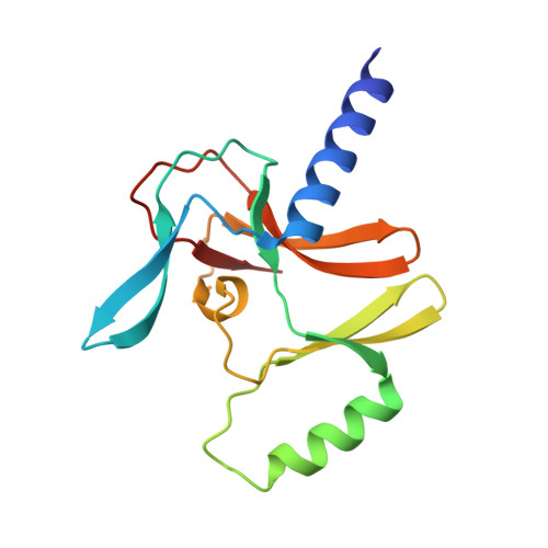
    Selective inhibitors of protein lysine methyltransferases, including SET domain-containing protein 8 (SETD8), are highly desired, as only a fraction of these enzymes are associated with high-quality inhibitors. From our previously discovered SETD8 inhibitor, we developed a more potent analog and solved a cocrystal structure, which is the first crystal structure of SETD8 in complex with a small-molecule inhibitor. This cocrystal structure allowed the design of a covalent inhibitor of SETD8 (MS453), which specifically modifies a cysteine residue near the inhibitor binding site, has an IC 50 value of 804 nM, reacts with SETD8 with near-quantitative yield, and is selective for SETD8 against 28 other methyltransferases. We also solved the crystal structure of the covalent inhibitor in complex with SETD8. This work provides atomic-level perspective on the inhibition of SETD8 by small molecules and will help identify high-quality chemical probes of SETD8.


  • Organizational Affiliation
    • Department of Pharmacological Sciences and Department of Oncological Sciences, Icahn School of Medicine at Mount Sinai , New York, New York 10029, United States.

Macromolecule Content 

  • Total Structure Weight: 17.22 kDa 
  • Atom Count: 1,145 
  • Modeled Residue Count: 143 
  • Deposited Residue Count: 148 
  • Unique protein chains: 1

Macromolecules

Find similar proteins by:|  3D Structure
Entity ID: 1
MoleculeChains  Sequence LengthOrganismDetailsImage
N-lysine methyltransferase KMT5A148Homo sapiensMutation(s): 1 
Gene Names: KMT5APRSET7SET07SET8SETD8
EC: 2.1.1 (PDB Primary Data), 2.1.1.43 (PDB Primary Data), 2.1.1.361 (UniProt)
UniProt & NIH Common Fund Data Resources
Find proteins for Q9NQR1 (Homo sapiens)
Explore Q9NQR1 
Go to UniProtKB:  Q9NQR1
PHAROS:  Q9NQR1
GTEx:  ENSG00000183955 
Entity Groups
Sequence Clusters30% Identity50% Identity70% Identity90% Identity95% Identity100% Identity
UniProt GroupQ9NQR1
Sequence Annotations
Expand
Reference Sequence

Small Molecules

Ligands 2 Unique
IDChains Name / Formula / InChI Key2D Diagram3D Interactions
75P

Query on 75P


Download:Ideal Coordinates CCD File
H [auth A]7-(2-aminoethoxy)-6-methoxy-2-(pyrrolidin-1-yl)-N-[5-(pyrrolidin-1-yl)pentyl]quinazolin-4-amine
C24 H38 N6 O2
UEDAXBZCRLISHC-UHFFFAOYSA-N
UNX

Query on UNX


Download:Ideal Coordinates CCD File
B [auth A]
C [auth A]
D [auth A]
E [auth A]
F [auth A]
B [auth A],
C [auth A],
D [auth A],
E [auth A],
F [auth A],
G [auth A]
UNKNOWN ATOM OR ION
X
Binding Affinity Annotations 
IDSourceBinding Affinity
75P BindingDB:  5T5G Kd: 1300 (nM) from 1 assay(s)
IC50: 1900 (nM) from 1 assay(s)

Experimental Data & Validation

Experimental Data

  • Method: X-RAY DIFFRACTION
  • Resolution: 2.10 Å
  • R-Value Free:  0.227 (Depositor), 0.237 (DCC) 
  • R-Value Work:  0.205 (Depositor), 0.214 (DCC) 
  • R-Value Observed: 0.206 (Depositor) 
Space Group: P 32 2 1
Unit Cell:
Length ( Å )Angle ( ˚ )
a = 88.957α = 90
b = 88.957β = 90
c = 34.989γ = 120
Software Package:
Software NamePurpose
REFMACrefinement
Aimlessdata scaling
PDB_EXTRACTdata extraction
XDSdata scaling
PHASERphasing

Structure Validation

View Full Validation Report


Ligand Structure Quality Assessment 


Entry History 

Revision History  (Full details and data files)

  • Version 1.0: 2016-09-28
    Type: Initial release
  • Version 1.1: 2016-10-26
    Changes: Derived calculations
  • Version 1.2: 2017-11-22
    Changes: Derived calculations, Refinement description
  • Version 1.3: 2018-05-23
    Changes: Data collection, Database references
  • Version 1.4: 2023-10-04
    Changes: Data collection, Database references, Refinement description
  • Version 1.5: 2024-10-16
    Changes: Structure summary