X-ray structures of fructosyl peptide oxidases revealing residues responsible for gating oxygen access in the oxidative half reaction
Shimasaki, T., Yoshida, H., Kamitori, S., Sode, K.(2017) Sci Rep 7: 2790-2790
- PubMed: 28584265 Search on PubMedSearch on PubMed Central
- DOI: https://doi.org/10.1038/s41598-017-02657-5
- Primary Citation Related Structures:
5T1E, 5T1F, 5XAO - PubMed Abstract:
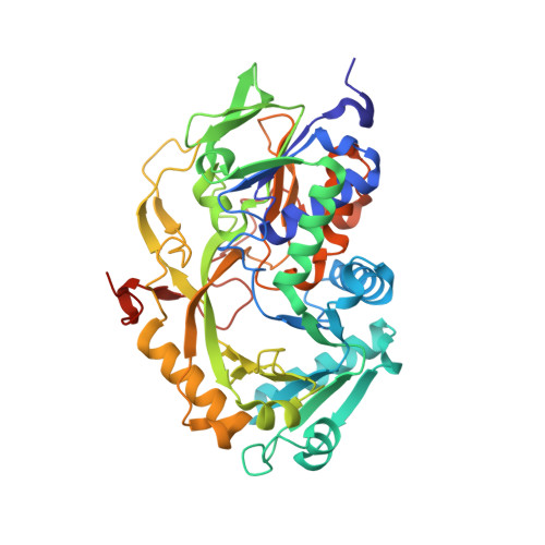
Current enzymatic systems for quantifying glycated hemoglobin are based on the FAD-containing enzyme fructosyl peptide oxidase (FPOX). FPOX has substrate specificity for fructosyl- α N-valyl-histidine derived from proteolytic digestion of the N-terminus of the HbA1c β-chain. This study reports the X-ray structures of the wild-type and Asn56Ala (N56A) mutant of Phaeosphaeria nodorum fructosyl peptide oxidase (PnFPOX) to elucidate the residues responsible for the oxidative half-reaction. N56A showed decreased oxidase activity compared to the wild -type, while its dye-mediated dehydrogenase activity was higher than that of wild type. In wild-type PnFPOX, Asn56 forms a hydrogen bond with Lys274, thereby preventing it from forming a salt bridge with Asp54. By contrast, Lys274 of PnFPOX N56A moves toward Asp54, and they approach each other to form a salt bridge at a distance of 2.92-3.35 Å. Site-directed mutagenesis studies and protein channel analysis suggest that Asp54 assists in accepting oxygen properly at the position of the bound water molecule in the main oxygen channel. These results reveal that Asn56 in PnFPOX is essential for maintaining an effective oxygen accession path, and support the role of Asp54 as a gate keeper that cooperates with Lys274 to enable oxygen to reach the active site properly.
- Department of Biotechnology and Life Science, Graduate School of Engineering, Tokyo University of Agriculture and Technology, 2-24-16, Nakamachi, Koganei, Tokyo, 184-8588, Japan.
Organizational Affiliation: