


Crystal Structure of a human phosphodiesterase 10 complex
Flohr, A., Schlatter, D., Kuhn, B., Rudolph, M.G.To be published.
Experimental Data Snapshot
Starting Model: other
View more details
Entity ID: 1 | |||||
|---|---|---|---|---|---|
| Molecule | Chains | Sequence Length | Organism | Details | Image |
| cAMP and cAMP-inhibited cGMP 3',5'-cyclic phosphodiesterase 10A | 343 | Homo sapiens | Mutation(s): 0 Gene Names: PDE10A EC: 3.1.4.17 | 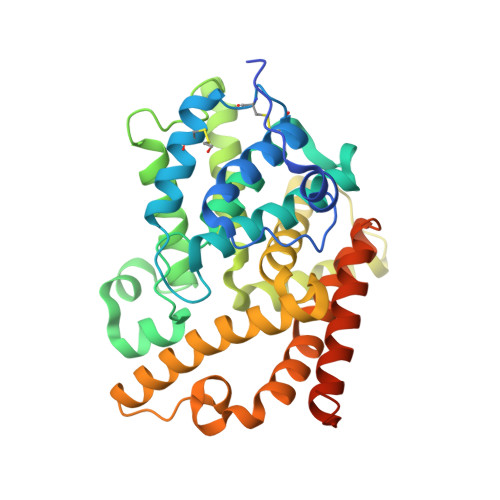 | |
UniProt & NIH Common Fund Data Resources | |||||
PHAROS: Q9Y233 GTEx: ENSG00000112541 | |||||
Entity Groups | |||||
| Sequence Clusters | 30% Identity50% Identity70% Identity90% Identity95% Identity100% Identity | ||||
| UniProt Group | Q9Y233 | ||||
Sequence AnnotationsExpand | |||||
Reference Sequence | |||||
| Ligands 4 Unique | |||||
|---|---|---|---|---|---|
| ID | Chains | Name / Formula / InChI Key | 2D Diagram | 3D Interactions | |
| KCF (Subject of Investigation/LOI) Download:Ideal Coordinates CCD File | G [auth A], K [auth B], O [auth C], R [auth D] | 4-bromanyl-~{N},~{N}-dimethyl-1$l^{4},7,9-triazabicyclo[4.3.0]nona-1,3,5-trien-8-amine C8 H9 Br N4 JATIVHGFJAULAI-UHFFFAOYSA-N |  | ||
| BR Download:Ideal Coordinates CCD File | H [auth A], L [auth B] | BROMIDE ION Br CPELXLSAUQHCOX-UHFFFAOYSA-M |  | ||
| ZN Download:Ideal Coordinates CCD File | E [auth A], I [auth B], M [auth C], P [auth D] | ZINC ION Zn PTFCDOFLOPIGGS-UHFFFAOYSA-N |  | ||
| MG Download:Ideal Coordinates CCD File | F [auth A], J [auth B], N [auth C], Q [auth D] | MAGNESIUM ION Mg JLVVSXFLKOJNIY-UHFFFAOYSA-N |  | ||
| Modified Residues 1 Unique | |||||
|---|---|---|---|---|---|
| ID | Chains | Type | Formula | 2D Diagram | Parent |
| CME Query on CME | A, B, C, D | L-PEPTIDE LINKING | C5 H11 N O3 S2 |  | CYS |
| Length ( Å ) | Angle ( ˚ ) |
|---|---|
| a = 135 | α = 90 |
| b = 135 | β = 90 |
| c = 234.995 | γ = 120 |
| Software Name | Purpose |
|---|---|
| XSCALE | data scaling |
| REFMAC | refinement |
| PDB_EXTRACT | data extraction |
| XDS | data reduction |
| XSCALE | data scaling |
| PHASER | phasing |
| Funding Organization | Location | Grant Number |
|---|---|---|
| F. Hoffmann-La Roche | Switzerland | -- |


