


Miniaturization process reloaded - structural and functional insights from a miniaturized rubredoxin
Chino, M., Di Costanzo, L.To be published.
Experimental Data Snapshot
Starting Model: in silico
View more details
wwPDB Validation 3D Report Full Report

Entity ID: 1 | |||||
|---|---|---|---|---|---|
| Molecule | Chains | Sequence Length | Organism | Details | Image |
| METP, miniaturized rubredoxin | A [auth C] | 30 | synthetic construct | Mutation(s): 0 | 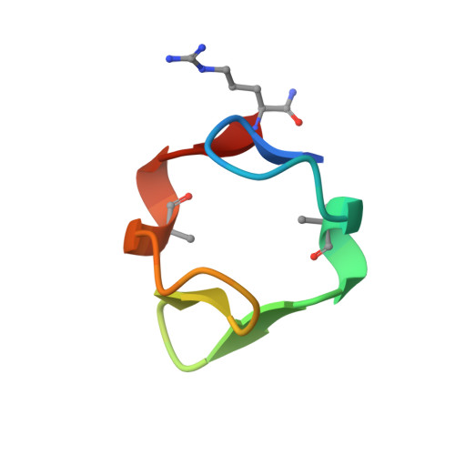 |
| Ligands 1 Unique | |||||
|---|---|---|---|---|---|
| ID | Chains | Name / Formula / InChI Key | 2D Diagram | 3D Interactions | |
| CD (Subject of Investigation/LOI) Download:Ideal Coordinates CCD File | B [auth C] | CADMIUM ION Cd WLZRMCYVCSSEQC-UHFFFAOYSA-N |  | ||
| Modified Residues 1 Unique | |||||
|---|---|---|---|---|---|
| ID | Chains | Type | Formula | 2D Diagram | Parent |
| AIB Query on AIB | A [auth C] | L-PEPTIDE LINKING | C4 H9 N O2 |  | ALA |
| Length ( Å ) | Angle ( ˚ ) |
|---|---|
| a = 37.07 | α = 90 |
| b = 56.654 | β = 90 |
| c = 19.331 | γ = 90 |
| Software Name | Purpose |
|---|---|
| PHENIX | refinement |
| PDB_EXTRACT | data extraction |
| XDS | data reduction |
| XDS | data scaling |
| PHENIX | phasing |


