5S9U

PanDDA analysis group deposition -- Crystal Structure of Trypanosoma brucei Trypanothione reductase in complex with Z32327641


Experimental Data Snapshot

  • Method: X-RAY DIFFRACTION
  • Resolution: 1.73 Å
  • R-Value Free: 0.229 
  • R-Value Work: 0.192 
  • R-Value Observed: 0.193 

wwPDB Validation   3D Report Full Report


Ligand Structure Quality Assessment 


This is version 1.1 of the entry. See complete history


Literature

Innovative Approach for a Classic Target: Fragment Screening on Trypanothione Reductase Reveals New Opportunities for Drug Design.

Fiorillo, A.Colotti, G.Exertier, C.Liuzzi, A.Seghetti, F.Salerno, A.Caciolla, J.Ilari, A.

(2022) Front Mol Biosci 9: 900882-900882

  • DOI: https://doi.org/10.3389/fmolb.2022.900882
  • Primary Citation of Related Structures:  
    5S9S, 5S9T, 5S9U, 5S9V, 5S9W, 5S9X, 5S9Y, 5S9Z, 5SA0, 5SA1, 5SA2, 5SA3, 5SMJ

  • PubMed Abstract: 

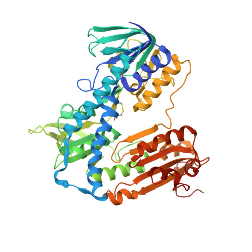
    Trypanothione reductase (TR) is a key factor in the redox homeostasis of trypanosomatid parasites, critical for survival in the hostile oxidative environment generated by the host to fight infection. TR is considered an attractive target for the development of new trypanocidal agents as it is essential for parasite survival but has no close homolog in humans. However, the high efficiency and turnover of TR challenging targets since only potent inhibitors, with nanomolar IC50, can significantly affect parasite redox state and viability. To aid the design of effective compounds targeting TR, we performed a fragment-based crystal screening at the Diamond Light Source XChem facility using a library optimized for follow-up synthesis steps. The experiment, allowing for testing over 300 compounds, resulted in the identification of 12 new ligands binding five different sites. Interestingly, the screening revealed the existence of an allosteric pocket close to the NADPH binding site, named the "doorstop pocket" since ligands binding at this site interfere with TR activity by hampering the "opening movement" needed to allow cofactor binding. The second remarkable site, known as the Z-site, identified by the screening, is located within the large trypanothione cavity but corresponds to a region not yet exploited for inhibition. The fragments binding to this site are close to each other and have some remarkable features making them ideal for follow-up optimization as a piperazine moiety in three out of five fragments.


  • Organizational Affiliation

    Institute of Molecular Biology and Pathology, Italian National Research Council, IBPM-CNR, Rome, Italy.


Macromolecules
Find similar proteins by:  (by identity cutoff)  |  3D Structure
Entity ID: 1
MoleculeChains Sequence LengthOrganismDetailsImage
N(1),N(8)-bis(glutathionyl)spermidine reductase
A, B
495Trypanosoma bruceiMutation(s): 0 
EC: 1.8.1.12
UniProt
Find proteins for Q389T8 (Trypanosoma brucei brucei (strain 927/4 GUTat10.1))
Explore Q389T8 
Go to UniProtKB:  Q389T8
Entity Groups  
Sequence Clusters30% Identity50% Identity70% Identity90% Identity95% Identity100% Identity
UniProt GroupQ389T8
Sequence Annotations
Expand
  • Reference Sequence
Small Molecules
Ligands 5 Unique
IDChains Name / Formula / InChI Key2D Diagram3D Interactions
FAD
Query on FAD

Download Ideal Coordinates CCD File 
C [auth A],
L [auth B]
FLAVIN-ADENINE DINUCLEOTIDE
C27 H33 N9 O15 P2
VWWQXMAJTJZDQX-UYBVJOGSSA-N
JMM (Subject of Investigation/LOI)
Query on JMM

Download Ideal Coordinates CCD File 
J [auth A],
Q [auth B]
[4-(cyclopropanecarbonyl)piperazin-1-yl](furan-2-yl)methanone
C13 H16 N2 O3
SMBREKYBPARCFW-UHFFFAOYSA-N
BR
Query on BR

Download Ideal Coordinates CCD File 
R [auth B]BROMIDE ION
Br
CPELXLSAUQHCOX-UHFFFAOYSA-M
DMS
Query on DMS

Download Ideal Coordinates CCD File 
D [auth A]
E [auth A]
F [auth A]
G [auth A]
H [auth A]
D [auth A],
E [auth A],
F [auth A],
G [auth A],
H [auth A],
I [auth A],
M [auth B],
N [auth B],
O [auth B],
P [auth B]
DIMETHYL SULFOXIDE
C2 H6 O S
IAZDPXIOMUYVGZ-UHFFFAOYSA-N
MG
Query on MG

Download Ideal Coordinates CCD File 
K [auth A],
S [auth B]
MAGNESIUM ION
Mg
JLVVSXFLKOJNIY-UHFFFAOYSA-N
Experimental Data & Validation

Experimental Data

  • Method: X-RAY DIFFRACTION
  • Resolution: 1.73 Å
  • R-Value Free: 0.229 
  • R-Value Work: 0.192 
  • R-Value Observed: 0.193 
  • Space Group: P 21 21 21
Unit Cell:
Length ( Å )Angle ( ˚ )
a = 79.96α = 90
b = 108.51β = 90
c = 111.68γ = 90
Software Package:
Software NamePurpose
REFMACrefinement
Aimlessdata scaling
PDB_EXTRACTdata extraction
XDSdata reduction
REFMACphasing

Structure Validation

View Full Validation Report


Ligand Structure Quality Assessment 


Entry History & Funding Information

Deposition Data


Funding OrganizationLocationGrant Number
iNEXT-DiscoveryEuropean UnionLB25167
CNCCSItalyB56G1500114000

Revision History  (Full details and data files)

  • Version 1.0: 2022-06-15
    Type: Initial release
  • Version 1.1: 2023-03-01
    Changes: Database references