5S5L | pdb_00005s5l

Tubulin-Z1492796719-complex


Experimental Data Snapshot

  • Method: X-RAY DIFFRACTION
  • Resolution: 2.25 Å
  • R-Value Free: 
    0.235 (Depositor), 0.236 (DCC) 
  • R-Value Work: 
    0.204 (Depositor), 0.207 (DCC) 
  • R-Value Observed: 
    0.206 (Depositor) 

Starting Model: experimental
View more details

wwPDB Validation 3D Report Full Report

Validation slider image for 5S5L

Ligand Structure Quality Assessment 


This is version 1.2 of the entry. See complete history

Literature

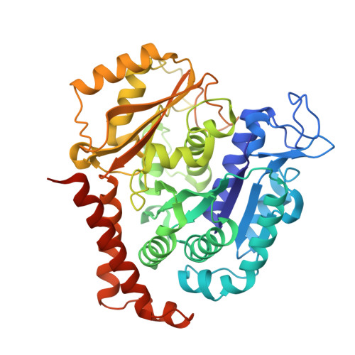
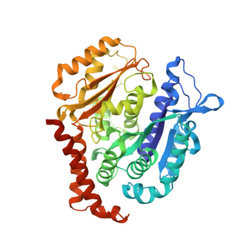
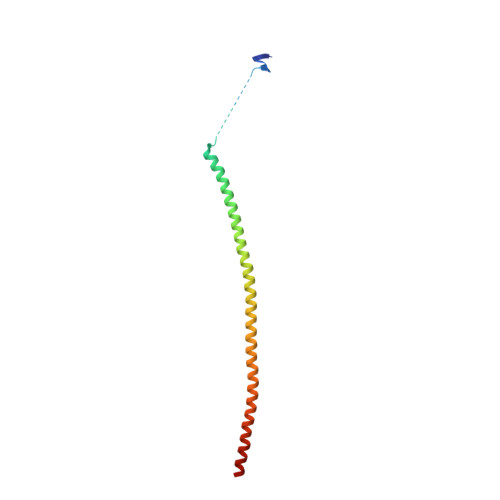
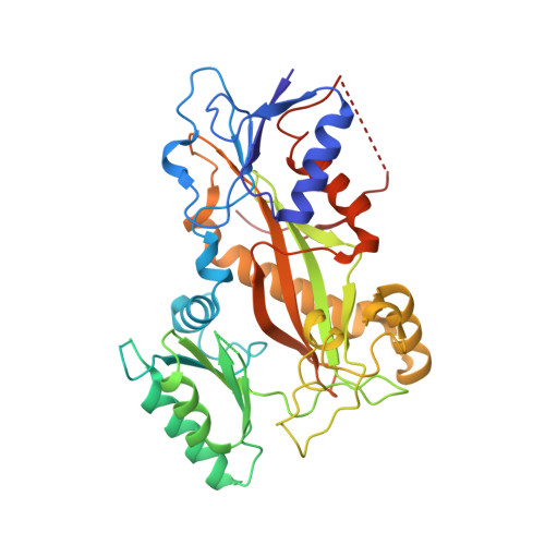
Comprehensive Analysis of Binding Sites in Tubulin.

Muhlethaler, T.Gioia, D.Prota, A.E.Sharpe, M.E.Cavalli, A.Steinmetz, M.O.

(2021) Angew Chem Int Ed Engl 60: 13331-13342

  • DOI: https://doi.org/10.1002/anie.202100273
  • Primary Citation Related Structures: 
    5S4L, 5S4M, 5S4N, 5S4O, 5S4P, 5S4Q, 5S4R, 5S4S, 5S4T, 5S4U, 5S4V, 5S4W, 5S4X, 5S4Y, 5S4Z, 5S50, 5S51, 5S52, 5S53, 5S54, 5S55, 5S56, 5S57, 5S58, 5S59, 5S5A, 5S5B, 5S5C, 5S5D, 5S5E, 5S5F, 5S5G, 5S5H, 5S5I, 5S5J, 5S5K, 5S5L, 5S5M, 5S5N, 5S5O, 5S5P, 5S5Q, 5S5R, 5S5S, 5S5T, 5S5U, 5S5V, 5S5W, 5S5X, 5S5Y, ... Search all related entries

  • PubMed Abstract: 

    Tubulin plays essential roles in vital cellular activities and is the target of a wide range of proteins and ligands. Here, using a combined computational and crystallographic fragment screening approach, we addressed the question of how many binding sites exist in tubulin. We identified 27 distinct sites, of which 11 have not been described previously, and analyzed their relationship to known tubulin-protein and tubulin-ligand interactions. We further observed an intricate pocket communication network and identified 56 chemically diverse fragments that bound to 10 distinct tubulin sites. Our results offer a unique structural basis for the development of novel small molecules for use as tubulin modulators in basic research applications or as drugs. Furthermore, our method lays down a framework that may help to discover new pockets in other pharmaceutically important targets and characterize them in terms of chemical tractability and allosteric modulation.


  • Organizational Affiliation
    • Laboratory of Biomolecular Research, Department of Biology and Chemistry, Paul Scherrer Institut, 5232, Villigen PSI, Switzerland.

Macromolecule Content 

  • Total Structure Weight: 264.74 kDa 
  • Atom Count: 18,052 
  • Modeled Residue Count: 2,203 
  • Deposited Residue Count: 2,319 
  • Unique protein chains: 4

Macromolecules

Find similar proteins by:|  3D Structure
Entity ID: 1
MoleculeChains  Sequence LengthOrganismDetailsImage
Tubulin alpha-1B chain
A, C
451Bos taurusMutation(s): 0 
EC: 3.6.5
UniProt
Find proteins for P81947 (Bos taurus)
Explore P81947 
Go to UniProtKB:  P81947
Entity Groups
Sequence Clusters30% Identity50% Identity70% Identity90% Identity95% Identity100% Identity
UniProt GroupP81947
Sequence Annotations
Expand
Reference Sequence
Find similar proteins by:|  3D Structure
Entity ID: 2
MoleculeChains  Sequence LengthOrganismDetailsImage
Tubulin beta-2B chain
B, D
445Bos taurusMutation(s): 0 
UniProt
Find proteins for Q6B856 (Bos taurus)
Explore Q6B856 
Go to UniProtKB:  Q6B856
Entity Groups
Sequence Clusters30% Identity50% Identity70% Identity90% Identity95% Identity100% Identity
UniProt GroupQ6B856
Sequence Annotations
Expand
Reference Sequence
Find similar proteins by:|  3D Structure
Entity ID: 3
MoleculeChains  Sequence LengthOrganismDetailsImage
Stathmin-4143Rattus norvegicusMutation(s): 0 
Gene Names: Stmn4
UniProt
Find proteins for P63043 (Rattus norvegicus)
Explore P63043 
Go to UniProtKB:  P63043
Entity Groups
Sequence Clusters30% Identity50% Identity70% Identity90% Identity95% Identity100% Identity
UniProt GroupP63043
Sequence Annotations
Expand
Reference Sequence
Find similar proteins by:|  3D Structure
Entity ID: 4
MoleculeChains  Sequence LengthOrganismDetailsImage
Tubulin-Tyrosine Ligase384Gallus gallusMutation(s): 0 
Gene Names: TTL

Small Molecules

Ligands 7 Unique
IDChains Name / Formula / InChI Key2D Diagram3D Interactions
GTP

Query on GTP


Download:Ideal Coordinates CCD File
G [auth A],
P [auth C]
GUANOSINE-5'-TRIPHOSPHATE
C10 H16 N5 O14 P3
XKMLYUALXHKNFT-UUOKFMHZSA-N
ACP

Query on ACP


Download:Ideal Coordinates CCD File
U [auth F]PHOSPHOMETHYLPHOSPHONIC ACID ADENYLATE ESTER
C11 H18 N5 O12 P3
UFZTZBNSLXELAL-IOSLPCCCSA-N
GDP

Query on GDP


Download:Ideal Coordinates CCD File
K [auth B],
S [auth D]
GUANOSINE-5'-DIPHOSPHATE
C10 H15 N5 O11 P2
QGWNDRXFNXRZMB-UUOKFMHZSA-N
MES

Query on MES


Download:Ideal Coordinates CCD File
N [auth B]2-(N-MORPHOLINO)-ETHANESULFONIC ACID
C6 H13 N O4 S
SXGZJKUKBWWHRA-UHFFFAOYSA-N
X0M
(Subject of Investigation/LOI)

Query on X0M


Download:Ideal Coordinates CCD File
O [auth B]N-[(3S)-1,2,3,4-tetrahydroquinolin-3-yl]acetamide
C11 H14 N2 O
HCEIEGOMGWEGOJ-JTQLQIEISA-N
CA

Query on CA


Download:Ideal Coordinates CCD File
I [auth A],
J [auth A],
M [auth B],
R [auth C]
CALCIUM ION
Ca
BHPQYMZQTOCNFJ-UHFFFAOYSA-N
MG

Query on MG


Download:Ideal Coordinates CCD File
H [auth A],
L [auth B],
Q [auth C],
T [auth D],
V [auth F]
MAGNESIUM ION
Mg
JLVVSXFLKOJNIY-UHFFFAOYSA-N

Experimental Data & Validation

Experimental Data

  • Method: X-RAY DIFFRACTION
  • Resolution: 2.25 Å
  • R-Value Free:  0.235 (Depositor), 0.236 (DCC) 
  • R-Value Work:  0.204 (Depositor), 0.207 (DCC) 
  • R-Value Observed: 0.206 (Depositor) 
Space Group: P 21 21 21
Unit Cell:
Length ( Å )Angle ( ˚ )
a = 105.12α = 90
b = 159.02β = 90
c = 179.19γ = 90
Software Package:
Software NamePurpose
PHENIXrefinement
XSCALEdata scaling
PDB_EXTRACTdata extraction
XDSdata reduction
PHENIXphasing

Structure Validation

View Full Validation Report


Ligand Structure Quality Assessment 


Entry History 

& Funding Information

Deposition Data


Funding OrganizationLocationGrant Number
iNEXT/Horizon 2020European UnionPID2692
NEON/Regione LombardiaItalyID239047
Swiss National Science FoundationSwitzerland31003A_166608
Swiss National Science FoundationSwitzerland31030A_192566

Revision History  (Full details and data files)

  • Version 1.0: 2021-06-30
    Type: Initial release
  • Version 1.1: 2024-03-06
    Changes: Advisory, Data collection, Database references
  • Version 1.2: 2026-02-18
    Changes: Refinement description, Structure summary