


PanDDA analysis group deposition
Schuller, M., Talon, R., Krojer, T., Brandao-Neto, J., Douangamath, A., Zhang, R., von Delft, F., Schuler, H., Kessler, B., Knapp, S., Bountra, C., Arrowsmith, C.H., Edwards, A., Elkins, J.To be published.
Experimental Data Snapshot
Starting Model: experimental
View more details
Entity ID: 1 | |||||
|---|---|---|---|---|---|
| Molecule | Chains | Sequence Length | Organism | Details | Image |
| Poly [ADP-ribose] polymerase 14 | 183 | Homo sapiens | Mutation(s): 0 Gene Names: PARP14, BAL2, KIAA1268 EC: 2.4.2.30 (PDB Primary Data), 2.4.2 (UniProt) | 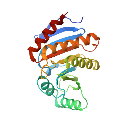 | |
UniProt & NIH Common Fund Data Resources | |||||
PHAROS: Q460N5 GTEx: ENSG00000173193 | |||||
Entity Groups | |||||
| Sequence Clusters | 30% Identity50% Identity70% Identity90% Identity95% Identity100% Identity | ||||
| UniProt Group | Q460N5 | ||||
Sequence AnnotationsExpand | |||||
Reference Sequence | |||||
| Ligands 3 Unique | |||||
|---|---|---|---|---|---|
| ID | Chains | Name / Formula / InChI Key | 2D Diagram | 3D Interactions | |
| AWP Download:Ideal Coordinates CCD File | E [auth A] | 1-cyclohexyl-3-(2-pyridin-4-ylethyl)urea C14 H21 N3 O XUPGANVQJLTRNJ-UHFFFAOYSA-N |  | ||
| DMS Download:Ideal Coordinates CCD File | D [auth A] | DIMETHYL SULFOXIDE C2 H6 O S IAZDPXIOMUYVGZ-UHFFFAOYSA-N |  | ||
| CL Download:Ideal Coordinates CCD File | B [auth A], C [auth A] | CHLORIDE ION Cl VEXZGXHMUGYJMC-UHFFFAOYSA-M |  | ||
| Length ( Å ) | Angle ( ˚ ) |
|---|---|
| a = 34.076 | α = 90 |
| b = 41.697 | β = 90 |
| c = 110.967 | γ = 90 |
| Software Name | Purpose |
|---|---|
| REFMAC | refinement |
| Aimless | data scaling |
| PDB_EXTRACT | data extraction |
| XDS | data reduction |
| REFMAC | phasing |


