


Crystal structure of Endothiapepsin
Huschmann, F.U., Weiss, M.S., Mueller, U., Haustedt, L.O., Klebe, G.To be published.
Experimental Data Snapshot
Entity ID: 1 | |||||
|---|---|---|---|---|---|
| Molecule | Chains | Sequence Length | Organism | Details | Image |
| Endothiapepsin | 330 | Cryphonectria parasitica | Mutation(s): 0 EC: 3.4.23.22 | 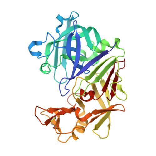 | |
UniProt | |||||
Entity Groups | |||||
| Sequence Clusters | 30% Identity50% Identity70% Identity90% Identity95% Identity100% Identity | ||||
| UniProt Group | P11838 | ||||
Sequence AnnotationsExpand | |||||
Reference Sequence | |||||
| Ligands 4 Unique | |||||
|---|---|---|---|---|---|
| ID | Chains | Name / Formula / InChI Key | 2D Diagram | 3D Interactions | |
| D6V Download:Ideal Coordinates CCD File | J [auth A] | 2-[(3S)-pyrrolidin-3-yl]-1H-benzimidazole C11 H13 N3 OFQGVINTJUCLFL-QMMMGPOBSA-N |  | ||
| GOL Download:Ideal Coordinates CCD File | B [auth A] C [auth A] D [auth A] E [auth A] F [auth A] | GLYCEROL C3 H8 O3 PEDCQBHIVMGVHV-UHFFFAOYSA-N |  | ||
| DMS Download:Ideal Coordinates CCD File | H [auth A], I [auth A] | DIMETHYL SULFOXIDE C2 H6 O S IAZDPXIOMUYVGZ-UHFFFAOYSA-N |  | ||
| ACT Download:Ideal Coordinates CCD File | K [auth A], L [auth A] | ACETATE ION C2 H3 O2 QTBSBXVTEAMEQO-UHFFFAOYSA-M |  | ||
| Length ( Å ) | Angle ( ˚ ) |
|---|---|
| a = 45.304 | α = 90 |
| b = 73.251 | β = 109.57 |
| c = 52.697 | γ = 90 |
| Software Name | Purpose |
|---|---|
| PHENIX | refinement |
| XSCALE | data scaling |
| PDB_EXTRACT | data extraction |


