


PanDDA analysis group deposition
Newman, J.A., Aitkenhead, H., Lee, S.Y., Kupinska, K., Burgess-Brown, N., Tallon, R., Krojer, T., von Delft, F., Arrowsmith, C.H., Edwards, A., Bountra, C., Gileadi, O.To be published.
Experimental Data Snapshot
Starting Model: experimental
View more details
wwPDB Validation 3D Report Full Report

Entity ID: 1 | |||||
|---|---|---|---|---|---|
| Molecule | Chains | Sequence Length | Organism | Details | Image |
| DCLRE1A | 343 | Homo sapiens | Mutation(s): 0 Gene Names: DCLRE1A, KIAA0086, SNM1, SNM1A EC: 3.5.2.6 | 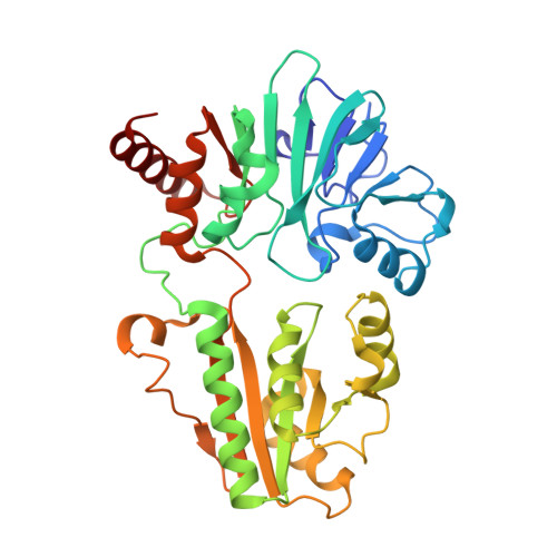 | |
UniProt & NIH Common Fund Data Resources | |||||
PHAROS: Q6PJP8 GTEx: ENSG00000198924 | |||||
Entity Groups | |||||
| Sequence Clusters | 30% Identity50% Identity70% Identity90% Identity95% Identity100% Identity | ||||
| UniProt Group | Q6PJP8 | ||||
Sequence AnnotationsExpand | |||||
Reference Sequence | |||||
| Ligands 2 Unique | |||||
|---|---|---|---|---|---|
| ID | Chains | Name / Formula / InChI Key | 2D Diagram | 3D Interactions | |
| MLI Download:Ideal Coordinates CCD File | B [auth A] | MALONATE ION C3 H2 O4 OFOBLEOULBTSOW-UHFFFAOYSA-L |  | ||
| NI Download:Ideal Coordinates CCD File | C [auth A] | NICKEL (II) ION Ni VEQPNABPJHWNSG-UHFFFAOYSA-N |  | ||
| Length ( Å ) | Angle ( ˚ ) |
|---|---|
| a = 51.68 | α = 90 |
| b = 57.11 | β = 90 |
| c = 114.38 | γ = 90 |
| Software Name | Purpose |
|---|---|
| REFMAC | refinement |
| Aimless | data scaling |
| PDB_EXTRACT | data extraction |
| XDS | data reduction |
| REFMAC | phasing |


