5OYJ

Crystal structure of VEGFR-2 domains 4-5 in complex with DARPin D4b


Experimental Data Snapshot

  • Method: X-RAY DIFFRACTION
  • Resolution: 2.38 Å
  • R-Value Free: 0.229 
  • R-Value Work: 0.178 
  • R-Value Observed: 0.180 

wwPDB Validation   3D Report Full Report


This is version 2.1 of the entry. See complete history


Literature

Characterization of a drug-targetable allosteric site regulating vascular endothelial growth factor signaling.

Thieltges, K.M.Avramovic, D.Piscitelli, C.L.Markovic-Mueller, S.Binz, H.K.Ballmer-Hofer, K.

(2018) Angiogenesis 21: 533-543

  • DOI: https://doi.org/10.1007/s10456-018-9606-9
  • Primary Citation of Related Structures:  
    5OYJ

  • PubMed Abstract: 

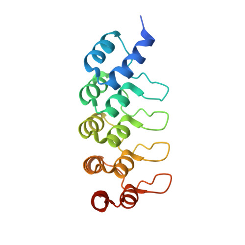
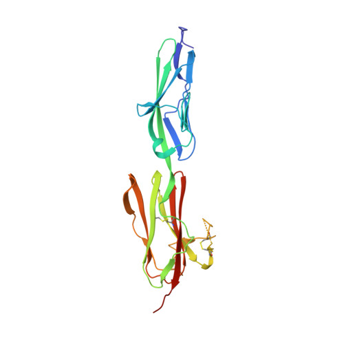
    Vascular endothelial growth factors (VEGFs) regulate blood and lymph vessel development upon activation of three receptor tyrosine kinases (VEGFRs). The extracellular domain of VEGFRs consists of seven Ig-homology domains, of which D2-3 form the ligand-binding site, while the membrane proximal domains D4-7 are involved in homotypic interactions in ligand-bound receptor dimers. Based on low-resolution structures, we identified allosteric sites in D4-5 and D7 of vascular endothelial growth factor receptor 2 (VEGFR-2) accomplishing regulatory functions. Allosteric inhibition of VEGFR-2 signaling represents an attractive option for the treatment of neovascular diseases. We showed earlier that DARPin ® binders to domains D4 or D7 are potent VEGFR-2 inhibitors. Here we investigated in detail the allosteric inhibition mechanism of the domain D4 binding inhibitor D4b. The 2.38 Å crystal structure of D4b in complex with VEGFR-2 D4-5, the first high-resolution structure of this VEGFR-2 segment, indicates steric hindrance by D4b as the mechanism of inhibition of receptor activation. At the cellular level, D4b triggered quantitative internalization of VEGFR-2 in the absence of ligand and thus clearance of VEGFR-2 from the surface of endothelial cells. The allosteric VEGFR-2 inhibition was sufficiently strong to efficiently inhibit the growth of human endothelial cells at suboptimal dose in a mouse xenograft model in vivo, underlining the therapeutic potential of the approach.


  • Organizational Affiliation

    Laboratory of Biomolecular Research, Paul Scherrer Institut, 5232, Villigen, Switzerland.


Macromolecules
Find similar proteins by:  (by identity cutoff)  |  3D Structure
Entity ID: 1
MoleculeChains Sequence LengthOrganismDetailsImage
DARPin D4b
A, B
169synthetic constructMutation(s): 0 
Entity Groups  
Sequence Clusters30% Identity50% Identity70% Identity90% Identity95% Identity100% Identity
Sequence Annotations
Expand
  • Reference Sequence
Find similar proteins by:  (by identity cutoff)  |  3D Structure
Entity ID: 2
MoleculeChains Sequence LengthOrganismDetailsImage
Vascular endothelial growth factor receptor 2
C, D
234Homo sapiensMutation(s): 0 
Gene Names: KDRFLK1VEGFR2
EC: 2.7.10.1
UniProt & NIH Common Fund Data Resources
Find proteins for P35968 (Homo sapiens)
Explore P35968 
Go to UniProtKB:  P35968
PHAROS:  P35968
GTEx:  ENSG00000128052 
Entity Groups  
Sequence Clusters30% Identity50% Identity70% Identity90% Identity95% Identity100% Identity
UniProt GroupP35968
Sequence Annotations
Expand
  • Reference Sequence
Oligosaccharides

Help

Entity ID: 3
MoleculeChains Length2D Diagram Glycosylation3D Interactions
2-acetamido-2-deoxy-beta-D-glucopyranose-(1-4)-2-acetamido-2-deoxy-beta-D-glucopyranose
E, F, G, H, I
E, F, G, H, I, J, K
2N-Glycosylation
Glycosylation Resources
GlyTouCan:  G42666HT
GlyCosmos:  G42666HT
GlyGen:  G42666HT
Entity ID: 4
MoleculeChains Length2D Diagram Glycosylation3D Interactions
alpha-D-glucopyranose-(1-1)-alpha-D-glucopyranose
L, M, N
2N/A
Glycosylation Resources
GlyTouCan:  G92130SN
GlyCosmos:  G92130SN
Small Molecules
Ligands 4 Unique
IDChains Name / Formula / InChI Key2D Diagram3D Interactions
CAC
Query on CAC

Download Ideal Coordinates CCD File 
T [auth C]CACODYLATE ION
C2 H6 As O2
OGGXGZAMXPVRFZ-UHFFFAOYSA-M
ACT
Query on ACT

Download Ideal Coordinates CCD File 
U [auth C]ACETATE ION
C2 H3 O2
QTBSBXVTEAMEQO-UHFFFAOYSA-M
CA
Query on CA

Download Ideal Coordinates CCD File 
O [auth A],
P [auth A],
Q [auth A],
S [auth C]
CALCIUM ION
Ca
BHPQYMZQTOCNFJ-UHFFFAOYSA-N
NA
Query on NA

Download Ideal Coordinates CCD File 
R [auth A]SODIUM ION
Na
FKNQFGJONOIPTF-UHFFFAOYSA-N
Biologically Interesting Molecules (External Reference) 1 Unique
Experimental Data & Validation

Experimental Data

  • Method: X-RAY DIFFRACTION
  • Resolution: 2.38 Å
  • R-Value Free: 0.229 
  • R-Value Work: 0.178 
  • R-Value Observed: 0.180 
  • Space Group: P 21 21 2
Unit Cell:
Length ( Å )Angle ( ˚ )
a = 109.489α = 90
b = 164.559β = 90
c = 70.759γ = 90
Software Package:
Software NamePurpose
PHENIXrefinement
XDSdata reduction
Aimlessdata scaling
PHASERphasing

Structure Validation

View Full Validation Report


Entry History & Funding Information

Deposition Data


Funding OrganizationLocationGrant Number
Switzerland--

Revision History  (Full details and data files)

  • Version 1.0: 2018-03-14
    Type: Initial release
  • Version 1.1: 2018-07-25
    Changes: Data collection, Database references
  • Version 2.0: 2020-07-29
    Type: Remediation
    Reason: Carbohydrate remediation
    Changes: Atomic model, Data collection, Derived calculations, Non-polymer description, Structure summary
  • Version 2.1: 2024-01-17
    Changes: Data collection, Database references, Refinement description, Structure summary