5OXS | pdb_00005oxs

Crystal structure of human lung surfactant protein D trimeric fragment with bound ligand Salmonella enterica Minnesota R5 oligosaccharide


Experimental Data Snapshot

  • Method: X-RAY DIFFRACTION
  • Resolution: 1.65 Å
  • R-Value Free: 
    0.187 (Depositor), 0.199 (DCC) 
  • R-Value Work: 
    0.165 (Depositor), 0.177 (DCC) 
  • R-Value Observed: 
    0.166 (Depositor) 

Starting Model: experimental
View more details

wwPDB Validation 3D Report Full Report

Validation slider image for 5OXS

This is version 2.2 of the entry. See complete history

Literature

Structural definition of hSP-D recognition of Salmonella enterica LPS inner core oligosaccharides reveals alternative binding modes for the same LPS.

Littlejohn, J.R.da Silva, R.F.Neale, W.A.Smallcombe, C.C.Clark, H.W.Mackay, R.A.Watson, A.S.Madsen, J.Hood, D.W.Burns, I.Greenhough, T.J.Shrive, A.K.

(2018) PLoS One 13: e0199175-e0199175

  • DOI: https://doi.org/10.1371/journal.pone.0199175
  • Primary Citation Related Structures: 
    5OXR, 5OXS

  • PubMed Abstract: 

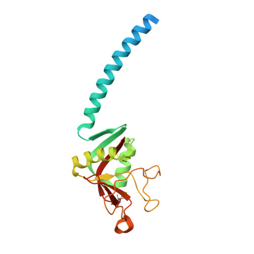
    The crystal structures of a biologically and therapeutically active recombinant homotrimeric fragment of native human SP-D (hSP-D) complexed with the inner core oligosaccharide of the Salmonella enterica sv Minnesota rough strains R5 and R7 (rough mutant chemotypes Rc and Rd1) have been determined. The structures reveal that hSP-D specifically and preferentially targets the LPS inner core via the innermost conserved Hep-Kdo pair with the flexibility for alternative recognition when this preferred epitope is not available for binding. Hep-Kdo binding is achieved through calcium dependent recognition of the heptose dihydroxyethyl side chain coupled with specific interactions between the Kdo and the binding site flanking residues Arg343 and Asp325 with evidence for an extended binding site for LPS inner cores containing multiple Kdo residues. In one subunit of the R5-bound structure this preferred mode of binding is precluded by the crystal lattice and oligosaccharide is bound through the terminal inner core glucose. The structures presented here thus provide unique multiple insights into the recognition and binding of bacterial LPS by hSP-D. Not only is it demonstrated that hSP-D targets the highly conserved LPS proximal inner core Hep-Kdo motif, but also that hSP-D can recognise either terminal or non-terminal sugars and has the flexibility and versatility to adopt alternative strategies for bacterial recognition, utilising alternative LPS epitopes when the preferred inner core Hep-Kdo disaccharide is not available for binding.


  • Organizational Affiliation
    • School of Life Sciences, Keele University, Staffordshire, United Kingdom.

Macromolecule Content 

  • Total Structure Weight: 59.01 kDa 
  • Atom Count: 4,175 
  • Modeled Residue Count: 458 
  • Deposited Residue Count: 531 
  • Unique protein chains: 1

Macromolecules

Find similar proteins by:|  3D Structure
Entity ID: 1
MoleculeChains  Sequence LengthOrganismDetailsImage
Pulmonary surfactant-associated protein D
A, B, C
177Homo sapiensMutation(s): 0 
Gene Names: SFTPDCOLEC7PSPDSFTP4
UniProt & NIH Common Fund Data Resources
Find proteins for P35247 (Homo sapiens)
Explore P35247 
Go to UniProtKB:  P35247
PHAROS:  P35247
GTEx:  ENSG00000133661 
Entity Groups
Sequence Clusters30% Identity50% Identity70% Identity90% Identity95% Identity100% Identity
UniProt GroupP35247
Sequence Annotations
Expand
Reference Sequence

Oligosaccharides

Help  
Entity ID: 2
MoleculeChains Length2D Diagram GlycosylationD Interactions
alpha-D-glucopyranose-(1-3)-L-glycero-alpha-D-manno-heptopyranose-(1-3)-L-glycero-alpha-D-manno-heptopyranose-(1-5)-4,7-anhydro-3-deoxy-D-gluco-oct-2-ulosonic acid
D, E, F
4N/A
Glycosylation Resources
GlyTouCan: G87317AK
GlyCosmos: G87317AK

Experimental Data & Validation

Experimental Data

  • Method: X-RAY DIFFRACTION
  • Resolution: 1.65 Å
  • R-Value Free:  0.187 (Depositor), 0.199 (DCC) 
  • R-Value Work:  0.165 (Depositor), 0.177 (DCC) 
  • R-Value Observed: 0.166 (Depositor) 
Space Group: P 1 21 1
Unit Cell:
Length ( Å )Angle ( ˚ )
a = 55.679α = 90
b = 108.51β = 92.89
c = 56.17γ = 90
Software Package:
Software NamePurpose
MOSFLMdata reduction
Aimlessdata scaling
REFMACrefinement
PDB_EXTRACTdata extraction

Structure Validation

View Full Validation Report


Entry History 

Deposition Data

Revision History  (Full details and data files)

  • Version 1.0: 2018-06-20
    Type: Initial release
  • Version 1.1: 2018-06-27
    Changes: Data collection, Database references
  • Version 2.0: 2020-07-29
    Type: Remediation
    Reason: Carbohydrate remediation
    Changes: Atomic model, Data collection, Derived calculations, Structure summary
  • Version 2.1: 2024-01-17
    Changes: Data collection, Database references, Derived calculations, Refinement description, Structure summary
  • Version 2.2: 2024-10-16
    Changes: Structure summary