5OOK | pdb_00005ook

Structure of A. marina Phycocyanin contains overlapping isoforms


Experimental Data Snapshot

  • Method: X-RAY DIFFRACTION
  • Resolution: 2.10 Å
  • R-Value Free: 
    0.215 (Depositor), 0.223 (DCC) 
  • R-Value Work: 
    0.193 (Depositor), 0.203 (DCC) 
  • R-Value Observed: 
    0.194 (Depositor) 

Starting Model: experimental
View more details

wwPDB Validation 3D Report Full Report

Validation slider image for 5OOK

Ligand Structure Quality Assessment 


This is version 1.3 of the entry. See complete history

Literature

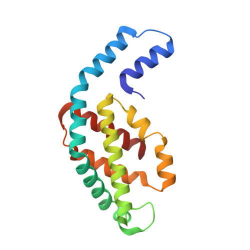
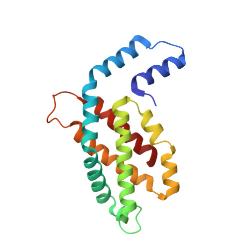
Structural heterogeneity leads to functional homogeneity in A. marina phycocyanin.

Bar-Zvi, S.Lahav, A.Harris, D.Niedzwiedzki, D.M.Blankenship, R.E.Adir, N.

(2018) Biochim Biophys Acta 1859: 544-553

  • DOI: https://doi.org/10.1016/j.bbabio.2018.04.007
  • Primary Citation Related Structures: 
    5OOK

  • PubMed Abstract: 

    The major light harvesting antenna in all cyanobacterial species is the phycobilisome (PBS). The smallest PBS identified to date is that of Acaryochloris marina (A. marina), composed of a single four-hexamer rod. We have determined the crystal structure of phycocyanin (AmPC), the major component of the A. marina PBS (AmPBS) to 2.1 Å. The basic unit of the AmPC is a heterodimer of two related subunits (α and β), and we show that the asymmetric unit contains a superposition of two α and two β isoforms, the products of the simultaneous expression of different genes. This is the first time to our knowledge that isolated proteins crystallized with such identifiable heterogeneity. We believe that the presence of the different isoforms allows the AmPBS to have a significant bathochromic shift in its fluorescence emission spectrum, allowing, in the total absence of allophycocyanin, a better overlap with absorption of the chlorophyll d-containing reaction centers. We show that this bathochromic shift exists in intact AmPBS as well as in its disassembled components, thus suggesting that AmPC can efficiently serve as the AmPBS terminal emitter.


  • Organizational Affiliation
    • Schulich Faculty of Chemistry, Technion-Israel Institute of Technology, Haifa 32000, Israel.

Macromolecule Content 

  • Total Structure Weight: 38.26 kDa 
  • Atom Count: 2,741 
  • Modeled Residue Count: 334 
  • Deposited Residue Count: 334 
  • Unique protein chains: 2

Macromolecules

Find similar proteins by:|  3D Structure
Entity ID: 1
MoleculeChains  Sequence LengthOrganismDetailsImage
Phycocyanin, alpha subunit162Acaryochloris marinaMutation(s): 0 
UniProt
Find proteins for A8ZMJ4 (Acaryochloris marina (strain MBIC 11017))
Explore A8ZMJ4 
Go to UniProtKB:  A8ZMJ4
Entity Groups
Sequence Clusters30% Identity50% Identity70% Identity90% Identity95% Identity100% Identity
UniProt GroupA8ZMJ4
Sequence Annotations
Expand
Reference Sequence
Find similar proteins by:|  3D Structure
Entity ID: 2
MoleculeChains  Sequence LengthOrganismDetailsImage
Phycocyanin, beta subunit172Acaryochloris marinaMutation(s): 0 
UniProt
Find proteins for A8ZMJ5 (Acaryochloris marina (strain MBIC 11017))
Explore A8ZMJ5 
Go to UniProtKB:  A8ZMJ5
Entity Groups
Sequence Clusters30% Identity50% Identity70% Identity90% Identity95% Identity100% Identity
UniProt GroupA8ZMJ5
Sequence Annotations
Expand
Reference Sequence

Small Molecules

Ligands 2 Unique
IDChains Name / Formula / InChI Key2D Diagram3D Interactions
CYC
(Subject of Investigation/LOI)

Query on CYC


Download:Ideal Coordinates CCD File
C [auth A],
H [auth B],
I [auth B]
PHYCOCYANOBILIN
C33 H40 N4 O6
VXTXPYZGDQPMHK-GMXXPEQVSA-N
PEG

Query on PEG


Download:Ideal Coordinates CCD File
D [auth A]
E [auth A]
F [auth A]
G [auth A]
J [auth B]
D [auth A],
E [auth A],
F [auth A],
G [auth A],
J [auth B],
K [auth B],
L [auth B],
M [auth B],
N [auth B],
O [auth B]
DI(HYDROXYETHYL)ETHER
C4 H10 O3
MTHSVFCYNBDYFN-UHFFFAOYSA-N

Experimental Data & Validation

Experimental Data

  • Method: X-RAY DIFFRACTION
  • Resolution: 2.10 Å
  • R-Value Free:  0.215 (Depositor), 0.223 (DCC) 
  • R-Value Work:  0.193 (Depositor), 0.203 (DCC) 
  • R-Value Observed: 0.194 (Depositor) 
Space Group: P 63
Unit Cell:
Length ( Å )Angle ( ˚ )
a = 152.806α = 90
b = 152.806β = 90
c = 39.312γ = 120
Software Package:
Software NamePurpose
REFMACrefinement
PHENIXrefinement
MOSFLMdata reduction
SCALAdata scaling
PHASERphasing

Structure Validation

View Full Validation Report


Ligand Structure Quality Assessment 


Entry History 

& Funding Information

Deposition Data


Funding OrganizationLocationGrant Number
United States - Israel Binational Science Foundation (BSF)Israel2014395

Revision History  (Full details and data files)

  • Version 1.0: 2018-06-20
    Type: Initial release
  • Version 1.1: 2022-03-30
    Changes: Author supporting evidence, Database references
  • Version 1.2: 2024-01-17
    Changes: Data collection, Refinement description
  • Version 1.3: 2024-11-13
    Changes: Structure summary