5OMI | pdb_00005omi

Crystal structure of GP2 from Lassa virus in a post fusion conformation


Experimental Data Snapshot

  • Method: X-RAY DIFFRACTION
  • Resolution: 2.56 Å
  • R-Value Free: 
    0.286 (Depositor), 0.293 (DCC) 
  • R-Value Work: 
    0.231 (Depositor), 0.240 (DCC) 
  • R-Value Observed: 
    0.237 (Depositor) 

Starting Model: experimental
View more details

wwPDB Validation 3D Report Full Report

Validation slider image for 5OMI

Ligand Structure Quality Assessment 


This is version 1.5 of the entry. See complete history

Literature

Variations in Core Packing of GP2 from Old World Mammarenaviruses in their Post-Fusion Conformations Affect Membrane-Fusion Efficiencies.

Shulman, A.Katz, M.Cohen-Dvashi, H.Greenblatt, H.M.Levy, Y.Diskin, R.

(2019) J Mol Biology 431: 2095-2111

  • DOI: https://doi.org/10.1016/j.jmb.2019.04.012
  • Primary Citation Related Structures: 
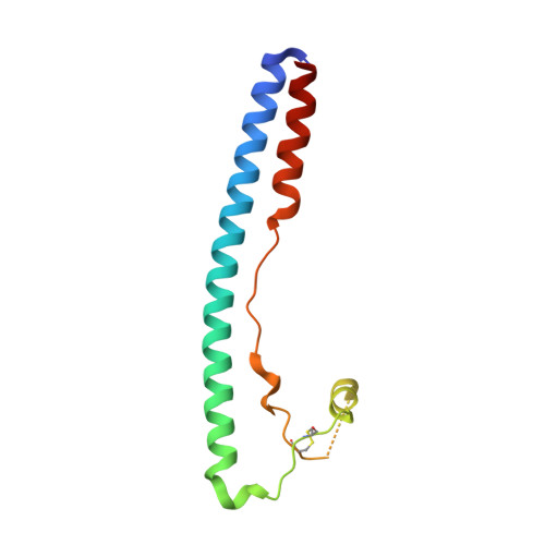
    5OMI

  • PubMed Abstract: 

    Lassa virus (LASV) is a notorious human pathogen in West Africa. Its class I trimeric spike complex displays a distinct architecture, and its cell entry mechanism involves unique attributes not shared by other related viruses. We determined the crystal structure of the GP2 fusion glycoprotein from the spike complex of LASV (GP2 LASV ) in its post-fusion conformation. GP2 LASV adopts a canonical helical bundle configuration similarly to other viruses in its family. The core packing of GP2 LASV , however, is more organized compared to GP2 from other viruses reducing the formation of internal hydrophobic cavities. We demonstrate a link between the formation of such unfavorable hydrophobic cavities and the efficiencies of membrane fusion and cell entry. Our study suggests that LASV has evolved a more efficient membrane fusogen compared to other viruses from its family by optimizing the post-fusion configuration of its GP2 module.


  • Organizational Affiliation
    • Department of Structural Biology, Weizmann Institute of Science, Rehovot, Israel.

Macromolecule Content 

  • Total Structure Weight: 41.95 kDa 
  • Atom Count: 2,714 
  • Modeled Residue Count: 325 
  • Deposited Residue Count: 354 
  • Unique protein chains: 1

Macromolecules

Find similar proteins by:|  3D Structure
Entity ID: 1
MoleculeChains  Sequence LengthOrganismDetailsImage
Pre-glycoprotein polyprotein GP complex
A, B, C
118Mammarenavirus lassaenseMutation(s): 1 
Gene Names: GPC
UniProt
Find proteins for P08669 (Lassa virus (strain Mouse/Sierra Leone/Josiah/1976))
Explore P08669 
Go to UniProtKB:  P08669
Entity Groups
Sequence Clusters30% Identity50% Identity70% Identity90% Identity95% Identity100% Identity
UniProt GroupP08669
Glycosylation
Glycosylation Sites: 1
Sequence Annotations
Expand
Reference Sequence

Experimental Data & Validation

Experimental Data

  • Method: X-RAY DIFFRACTION
  • Resolution: 2.56 Å
  • R-Value Free:  0.286 (Depositor), 0.293 (DCC) 
  • R-Value Work:  0.231 (Depositor), 0.240 (DCC) 
  • R-Value Observed: 0.237 (Depositor) 
Space Group: C 2 2 21
Unit Cell:
Length ( Å )Angle ( ˚ )
a = 58.318α = 90
b = 103.433β = 90
c = 171.551γ = 90
Software Package:
Software NamePurpose
PHENIXrefinement
XDSdata reduction
Aimlessdata scaling
PHASERphasing

Structure Validation

View Full Validation Report


Ligand Structure Quality Assessment 


Entry History 

& Funding Information

Deposition Data


Funding OrganizationLocationGrant Number
I-COREIsrael1775/12

Revision History  (Full details and data files)

  • Version 1.0: 2018-08-29
    Type: Initial release
  • Version 1.1: 2019-05-08
    Changes: Data collection, Database references
  • Version 1.2: 2019-06-05
    Changes: Data collection, Database references
  • Version 1.3: 2020-07-29
    Type: Remediation
    Reason: Carbohydrate remediation
    Changes: Data collection, Derived calculations, Structure summary
  • Version 1.4: 2024-01-17
    Changes: Data collection, Database references, Refinement description, Structure summary
  • Version 1.5: 2024-10-23
    Changes: Structure summary