5OLY | pdb_00005oly

5-fluorotryptophan labeled beta-phosphoglucomutase in a closed conformation, monoclinic crystal form


Experimental Data Snapshot

  • Method: X-RAY DIFFRACTION
  • Resolution: 2.00 Å
  • R-Value Free: 
    0.255 (Depositor), 0.262 (DCC) 
  • R-Value Work: 
    0.204 (Depositor), 0.218 (DCC) 
  • R-Value Observed: 
    0.207 (Depositor) 

Starting Model: experimental
View more details

wwPDB Validation 3D Report Full Report

Validation slider image for 5OLY

Ligand Structure Quality Assessment 


This is version 1.4 of the entry. See complete history

Literature

Observing enzyme ternary transition state analogue complexes by19F NMR spectroscopy.

Ampaw, A.Carroll, M.von Velsen, J.Bhattasali, D.Cohen, A.Bowler, M.W.Jakeman, D.L.

(2017) Chem Sci 8: 8427-8434

  • DOI: https://doi.org/10.1039/c7sc04204c
  • Primary Citation Related Structures: 
    5OLW, 5OLX, 5OLY

  • PubMed Abstract: 

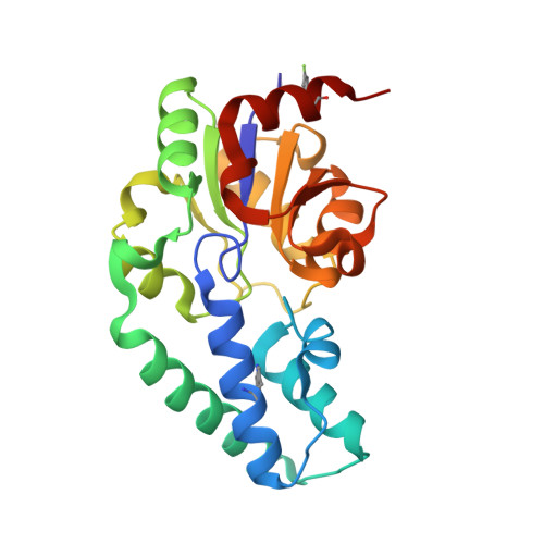
    Ternary transition state analogue (TSA) complexes probing the isomerization of β-d-glucose 1-phosphate (G1P) into d-glucose 6-phosphate (G6P) catalyzed by catalytically active, fluorinated (5-fluorotryptophan), β-phosphoglucomutase (βPGM) have been observed directly by 19 F NMR spectroscopy. In these complexes MgF 3 - and AlF 4 - are surrogates for the transferring phosphate. However, the relevance of these metal fluorides as TSA complexes has been queried. The 1D 19 F spectrum of a ternary TSA complex presented a molar equivalence between fluorinated enzyme, metal fluoride and non-isomerizable fluoromethylenephosphonate substrate analogue. Ring flips of the 5-fluoroindole ring remote from the active site were observed by both 19 F NMR and X-ray crystallography, but did not perturb function. This data unequivocally demonstrates that the concentration of the metal fluoride complexes is equivalent to the concentration of enzyme and ligand in the TSA complex in aqueous solution.


  • Organizational Affiliation
    • Department of Chemistry , Dalhousie University , Halifax , NS , Canada B3H 4R2 . Email: david.jakeman@dal.ca.

Macromolecule Content 

  • Total Structure Weight: 49.33 kDa 
  • Atom Count: 3,858 
  • Modeled Residue Count: 441 
  • Deposited Residue Count: 442 
  • Unique protein chains: 1

Macromolecules

Find similar proteins by:|  3D Structure
Entity ID: 1
MoleculeChains  Sequence LengthOrganismDetailsImage
Beta-phosphoglucomutaseA,
B [auth G]
221Lactococcus lactis subsp. lactisMutation(s): 0 
Gene Names: pgmBLL0429L0001
EC: 5.4.2.6
UniProt
Find proteins for P71447 (Lactococcus lactis subsp. lactis (strain IL1403))
Explore P71447 
Go to UniProtKB:  P71447
Entity Groups
Sequence Clusters30% Identity50% Identity70% Identity90% Identity95% Identity100% Identity
UniProt GroupP71447
Sequence Annotations
Expand
Reference Sequence

Small Molecules

Ligands 3 Unique
IDChains Name / Formula / InChI Key2D Diagram3D Interactions
G6P

Query on G6P


Download:Ideal Coordinates CCD File
E [auth A],
I [auth G]
6-O-phosphono-alpha-D-glucopyranose
C6 H13 O9 P
NBSCHQHZLSJFNQ-DVKNGEFBSA-N
MGF

Query on MGF


Download:Ideal Coordinates CCD File
C [auth A],
J [auth G]
TRIFLUOROMAGNESATE
F3 Mg
GJOMWUHGUQLOAC-UHFFFAOYSA-K
MG

Query on MG


Download:Ideal Coordinates CCD File
D [auth A],
F [auth A],
G,
H [auth G]
MAGNESIUM ION
Mg
JLVVSXFLKOJNIY-UHFFFAOYSA-N
Modified Residues  1 Unique
IDChains TypeFormula2D DiagramParent
FTR
Query on FTR
A,
B [auth G]
L-PEPTIDE LINKINGC11 H11 F N2 O2TRP

Experimental Data & Validation

Experimental Data

  • Method: X-RAY DIFFRACTION
  • Resolution: 2.00 Å
  • R-Value Free:  0.255 (Depositor), 0.262 (DCC) 
  • R-Value Work:  0.204 (Depositor), 0.218 (DCC) 
  • R-Value Observed: 0.207 (Depositor) 
Space Group: P 1 21 1
Unit Cell:
Length ( Å )Angle ( ˚ )
a = 57.737α = 90
b = 36.714β = 105.27
c = 107.853γ = 90
Software Package:
Software NamePurpose
PHENIXrefinement
XDSdata reduction
Aimlessdata scaling
MOLREPphasing

Structure Validation

View Full Validation Report


Ligand Structure Quality Assessment 


Entry History 

Deposition Data

Revision History  (Full details and data files)

  • Version 1.0: 2017-11-01
    Type: Initial release
  • Version 1.1: 2017-11-15
    Changes: Database references
  • Version 1.2: 2018-04-18
    Changes: Data collection, Database references
  • Version 1.3: 2020-07-29
    Type: Remediation
    Reason: Carbohydrate remediation
    Changes: Data collection, Derived calculations, Structure summary
  • Version 1.4: 2024-01-17
    Changes: Data collection, Database references, Refinement description, Structure summary