Structural basis for enzyme bifunctionality - the case of Gan1D from Geobacillus stearothermophilus.
Lansky, S., Zehavi, A., Belrhali, H., Shoham, Y., Shoham, G.(2017) FEBS J 284: 3931-3953
- PubMed: 28975708 Search on PubMed
- DOI: https://doi.org/10.1111/febs.14283
- Primary Citation Related Structures:
5OK7, 5OKA, 5OKB, 5OKE, 5OKG, 5OKH, 5OKJ, 5OKK, 5OKQ, 5OKR, 5OKS - PubMed Abstract:
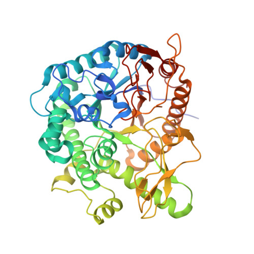
6-phospho-β-glucosidases and 6-phospho-β-galactosidases are enzymes that hydrolyze the β-glycosidic bond between a terminal non-reducing glucose-6-phosphate (Glc6P) or galactose-6-phosphate (Gal6P), respectively, and other organic molecules. Gan1D, a glycoside hydrolase (GH) belonging to the GH1 family, has recently been identified in a newly characterized galactan-utilization gene cluster in the bacterium Geobacillus stearothermophilus T-1. Gan1D has been shown to exhibit bifunctional activity, possessing both 6-phospho-β-galactosidase and 6-phospho-β-glucosidase activities. We report herein the complete 3D crystal structure of Gan1D, together with its acid/base catalytic mutant Gan1D-E170Q. The tertiary structure of Gan1D conforms well to the (β/α) 8 TIM-barrel fold commonly observed in GH enzymes, and its quaternary structure adopts a dimeric assembly, confirmed by gel-filtration and small-angle X-ray scattering results. We present also the structures of Gan1D in complex with the putative substrate cellobiose-6-phosphate (Cell6P) and the degradation products Glc6P and Gal6P. These complexes reveal the specific enzyme-substrate and enzyme-product binding interactions of Gan1D, and the residues involved in its glycone, aglycone, and phosphate binding sites. We show that the different ligands trapped in the active sites adopt different binding modes to the protein, providing a structural basis for the dual galactosidase/glucosidase activity observed for this enzyme. Based on this information, specific mutations were performed on one of the active site residues (W433), shifting the enzyme specificity from dual activity to a significant preference toward 6-phospho-β-glucosidase activity. These data and their comparison with structural data of related glucosidases and galactosidases are used for a more general discussion on the structure-function relationships in this sub-group of GH1 enzymes. Atomic coordinates of Gan1D-wild-type (WT)-P1, Gan1D-WT-C2, Gan1D-E170Q, Gan1D-WT-Gal6P, Gan1D-WT-Glc6P, and Gan1D-E170Q-Cell6P have been deposited in the Research Collaboratory for Structural Bioinformatics (RCSB) Protein Data Bank, under accession codes 5OKB, 5OKJ/5OKH, 5OKA/5OK7, 5OKQ/5OKK, 5OKS/5OKR, and 5OKG/5OKE, respectively.
- Institute of Chemistry, The Laboratory for Structural Chemistry and Biology, The Hebrew University of Jerusalem, Israel.
Organizational Affiliation: