5OFS | pdb_00005ofs

X-ray structure of a zinc binding GB1 mutant


Experimental Data Snapshot

  • Method: X-RAY DIFFRACTION
  • Resolution: 1.10 Å
  • R-Value Free: 
    0.145 (Depositor), 0.151 (DCC) 
  • R-Value Work: 
    0.125 (Depositor), 0.134 (DCC) 
  • R-Value Observed: 
    0.126 (Depositor) 

Starting Model: experimental
View more details

wwPDB Validation 3D Report Full Report

Validation slider image for 5OFS

This is version 1.2 of the entry. See complete history

Literature

Genetic Algorithm Based Design and Experimental Characterization of a Highly Thermostable Metalloprotein.

Bozkurt, E.Perez, M.A.S.Hovius, R.Browning, N.J.Rothlisberger, U.

(2018) J Am Chem Soc 140: 4517-4521

  • DOI: https://doi.org/10.1021/jacs.7b10660
  • Primary Citation Related Structures: 
    5O94, 5OFS

  • PubMed Abstract: 

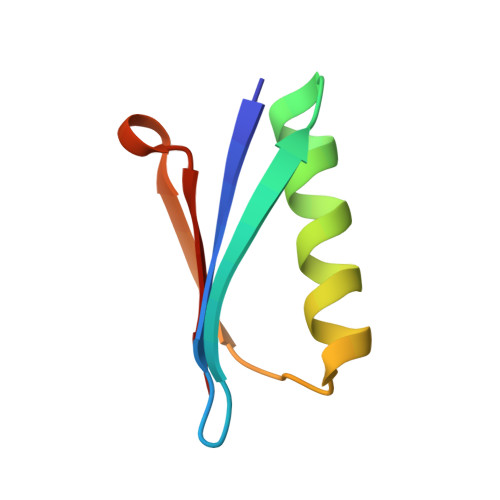
    The development of thermostable and solvent-tolerant metalloproteins is a long-sought goal for many applications in synthetic biology and biotechnology. In this work, we were able to engineer a highly thermostable and organic solvent-stable metallo variant of the B1 domain of protein G (GB1) with a tetrahedral zinc binding site reminiscent of the one of thermolysin. Promising candidates were designed computationally by applying a protocol based on classical and first-principles molecular dynamics simulations in combination with genetic algorithm optimization. The most promising of the computationally predicted mutants was expressed and structurally characterized and yielded a highly thermostable protein. The experimental results thus confirm the predictive power of the applied computational protein engineering approach for the de novo design of highly stable metalloproteins.


  • Organizational Affiliation
    • Laboratory of Computational Chemistry and Biochemistry , École Polytechnique Fédérale de Lausanne , CH-1015 Lausanne , Switzerland.

Macromolecule Content 

  • Total Structure Weight: 26.89 kDa 
  • Atom Count: 2,176 
  • Modeled Residue Count: 224 
  • Deposited Residue Count: 224 
  • Unique protein chains: 1

Macromolecules

Find similar proteins by:|  3D Structure
Entity ID: 1
MoleculeChains  Sequence LengthOrganismDetailsImage
Immunoglobulin G-binding protein G
A, B, C, D
56Streptococcus sp. 'group GMutation(s): 0 
Gene Names: spg

Small Molecules

Ligands 7 Unique
IDChains Name / Formula / InChI Key2D Diagram3D Interactions
MRD

Query on MRD


Download:Ideal Coordinates CCD File
Q [auth D](4R)-2-METHYLPENTANE-2,4-DIOL
C6 H14 O2
SVTBMSDMJJWYQN-RXMQYKEDSA-N
MPD

Query on MPD


Download:Ideal Coordinates CCD File
K [auth B],
M [auth C]
(4S)-2-METHYL-2,4-PENTANEDIOL
C6 H14 O2
SVTBMSDMJJWYQN-YFKPBYRVSA-N
SO4

Query on SO4


Download:Ideal Coordinates CCD File
T [auth D]SULFATE ION
O4 S
QAOWNCQODCNURD-UHFFFAOYSA-L
ZN
(Subject of Investigation/LOI)

Query on ZN


Download:Ideal Coordinates CCD File
E [auth A]
F [auth A]
J [auth B]
L [auth C]
O [auth D]
E [auth A],
F [auth A],
J [auth B],
L [auth C],
O [auth D],
P [auth D]
ZINC ION
Zn
PTFCDOFLOPIGGS-UHFFFAOYSA-N
EDO

Query on EDO


Download:Ideal Coordinates CCD File
I [auth A],
N [auth C],
V [auth D],
W [auth D],
X [auth D]
1,2-ETHANEDIOL
C2 H6 O2
LYCAIKOWRPUZTN-UHFFFAOYSA-N
ACT

Query on ACT


Download:Ideal Coordinates CCD File
G [auth A],
H [auth A],
U [auth D]
ACETATE ION
C2 H3 O2
QTBSBXVTEAMEQO-UHFFFAOYSA-M
CL

Query on CL


Download:Ideal Coordinates CCD File
R [auth D],
S [auth D]
CHLORIDE ION
Cl
VEXZGXHMUGYJMC-UHFFFAOYSA-M

Experimental Data & Validation

Experimental Data

  • Method: X-RAY DIFFRACTION
  • Resolution: 1.10 Å
  • R-Value Free:  0.145 (Depositor), 0.151 (DCC) 
  • R-Value Work:  0.125 (Depositor), 0.134 (DCC) 
  • R-Value Observed: 0.126 (Depositor) 
Space Group: P 21 21 2
Unit Cell:
Length ( Å )Angle ( ˚ )
a = 74.452α = 90
b = 75.621β = 90
c = 51.153γ = 90
Software Package:
Software NamePurpose
REFMACrefinement
ADDREFdata reduction
Aimlessdata scaling
REFMACphasing

Structure Validation

View Full Validation Report


Entry History 

& Funding Information

Deposition Data


Funding OrganizationLocationGrant Number
Swiss National Science FoundationSwitzerland200020-146645

Revision History  (Full details and data files)

  • Version 1.0: 2018-04-04
    Type: Initial release
  • Version 1.1: 2018-04-11
    Changes: Data collection, Database references, Derived calculations, Structure summary
  • Version 1.2: 2024-01-17
    Changes: Data collection, Database references, Derived calculations, Refinement description, Structure summary