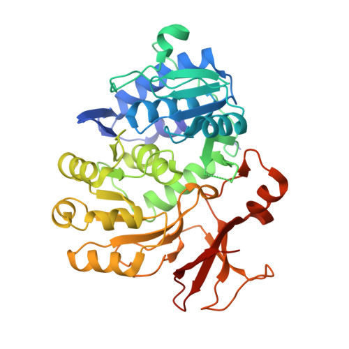
5OE5

Crystal structure of the N-terminal domain of PqsA in complex with anthraniloyl-AMP (crystal form 3)


Experimental Data Snapshot

  • Method: X-RAY DIFFRACTION
  • Resolution: 1.74 Å
  • R-Value Free: 0.277 
  • R-Value Work: 0.228 
  • R-Value Observed: 0.230 

wwPDB Validation   3D Report Full Report


Ligand Structure Quality Assessment 


This is version 1.2 of the entry. See complete history


Literature

Structures of the N-Terminal Domain of PqsA in Complex with Anthraniloyl- and 6-Fluoroanthraniloyl-AMP: Substrate Activation in Pseudomonas Quinolone Signal (PQS) Biosynthesis.

Witzgall, F.Ewert, W.Blankenfeldt, W.

(2017) Chembiochem 18: 2045-2055

  • DOI: https://doi.org/10.1002/cbic.201700374
  • Primary Citation of Related Structures:  
    5OE3, 5OE4, 5OE5, 5OE6

  • PubMed Abstract: 

    Pseudomonas aeruginosa, a prevalent pathogen in nosocomial infections and a major burden in cystic fibrosis, uses three interconnected quorum-sensing systems to coordinate virulence processes. At variance with other Gram-negative bacteria, one of these systems relies on 2-alkyl-4(1H)-quinolones (Pseudomonas quinolone signal, PQS) and might hence be an attractive target for new anti-infective agents. Here we report crystal structures of the N-terminal domain of anthranilate-CoA ligase PqsA, the first enzyme of PQS biosynthesis, in complex with anthraniloyl-AMP and with 6-fluoroanthraniloyl-AMP (6FABA-AMP) at 1.4 and 1.7 Å resolution. We find that PqsA belongs to an unrecognized subfamily of anthranilate-CoA ligases that recognize the amino group of anthranilate through a water-mediated hydrogen bond. The complex with 6FABA-AMP explains why 6FABA, an inhibitor of PQS biosynthesis, is a good substrate of PqsA. Together, our data might pave a way to new pathoblockers in P. aeruginosa infections.


  • Organizational Affiliation

    Structure and Function of Proteins, Helmholtz Centre for Infection Research, Inhoffenstrasse 7, 38124, Braunschweig, Germany.


Macromolecules
Find similar proteins by:  (by identity cutoff)  |  3D Structure
Entity ID: 1
MoleculeChains Sequence LengthOrganismDetailsImage
Anthranilate--CoA ligase407Pseudomonas aeruginosa PAO1Mutation(s): 0 
Gene Names: pqsAPA0996
EC: 6.2.1.32
UniProt
Find proteins for Q9I4X3 (Pseudomonas aeruginosa (strain ATCC 15692 / DSM 22644 / CIP 104116 / JCM 14847 / LMG 12228 / 1C / PRS 101 / PAO1))
Explore Q9I4X3 
Go to UniProtKB:  Q9I4X3
Entity Groups  
Sequence Clusters30% Identity50% Identity70% Identity90% Identity95% Identity100% Identity
UniProt GroupQ9I4X3
Sequence Annotations
Expand
  • Reference Sequence
Small Molecules
Ligands 2 Unique
IDChains Name / Formula / InChI Key2D Diagram3D Interactions
3UK
Query on 3UK

Download Ideal Coordinates CCD File 
B [auth A]5'-O-[(S)-[(2-aminobenzoyl)oxy](hydroxy)phosphoryl]adenosine
C17 H19 N6 O8 P
XZXXWUQOHYJTTC-XNIJJKJLSA-N
PEG
Query on PEG

Download Ideal Coordinates CCD File 
C [auth A],
D [auth A]
DI(HYDROXYETHYL)ETHER
C4 H10 O3
MTHSVFCYNBDYFN-UHFFFAOYSA-N
Experimental Data & Validation

Experimental Data

  • Method: X-RAY DIFFRACTION
  • Resolution: 1.74 Å
  • R-Value Free: 0.277 
  • R-Value Work: 0.228 
  • R-Value Observed: 0.230 
  • Space Group: P 21 21 2
Unit Cell:
Length ( Å )Angle ( ˚ )
a = 69.7α = 90
b = 84.204β = 90
c = 64.237γ = 90
Software Package:
Software NamePurpose
Aimlessdata scaling
PHASERphasing
PHENIXrefinement
PDB_EXTRACTdata extraction
XDSdata reduction
Cootmodel building

Structure Validation

View Full Validation Report


Ligand Structure Quality Assessment 


Entry History 

Deposition Data

Revision History  (Full details and data files)

  • Version 1.0: 2017-09-06
    Type: Initial release
  • Version 1.1: 2017-10-25
    Changes: Database references
  • Version 1.2: 2024-05-08
    Changes: Data collection, Database references