5OCP | pdb_00005ocp

The periplasmic binding protein component of the arabinose ABC transporter from Shewanella sp. ANA-3 bound to alpha and beta-L-arabinofuranose


Experimental Data Snapshot

  • Method: X-RAY DIFFRACTION
  • Resolution: 1.70 Å
  • R-Value Free: 
    0.204 (Depositor), 0.224 (DCC) 
  • R-Value Work: 
    0.167 (Depositor) 
  • R-Value Observed: 
    0.169 (Depositor) 

Starting Model: experimental
View more details

wwPDB Validation 3D Report Full Report

Validation slider image for 5OCP

Ligand Structure Quality Assessment 


This is version 3.2 of the entry. See complete history

Literature

CHARACTERISATION OF A FURANOSE SPECIFIC ABC TRANSPORTER ESSENTIAL FOR ARABINOSE UTILISATION FROM THE LIGNOCELLULOSE DEGRADING BACTERIUM SHEWANELLA SP. ANA-3

Herman, R.Drousiotis, K.Wilkinson, A.J.Thomas, G.H.

To be published.

Macromolecule Content 

  • Total Structure Weight: 67.01 kDa 
  • Atom Count: 5,171 
  • Modeled Residue Count: 604 
  • Deposited Residue Count: 604 
  • Unique protein chains: 1

Macromolecules

Find similar proteins by:|  3D Structure
Entity ID: 1
MoleculeChains  Sequence LengthOrganismDetailsImage
Periplasmic binding protein/LacI transcriptional regulator
A, B
302Shewanella sp. ANA-3Mutation(s): 0 
Gene Names: Shewana3_2073
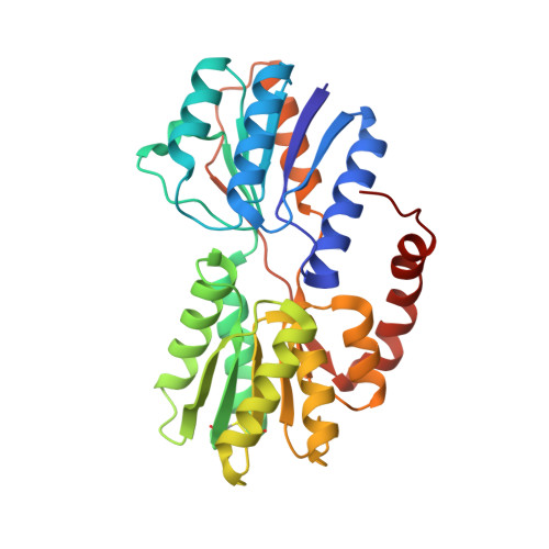
UniProt
Find proteins for A0ACD6B8D4 (Shewanella sp. (strain ANA-3))
Explore A0ACD6B8D4 
Go to UniProtKB:  A0ACD6B8D4
Entity Groups
Sequence Clusters30% Identity50% Identity70% Identity90% Identity95% Identity100% Identity
UniProt GroupA0ACD6B8D4
Sequence Annotations
Expand
Reference Sequence

Small Molecules

Ligands 4 Unique
IDChains Name / Formula / InChI Key2D Diagram3D Interactions
FUB
(Subject of Investigation/LOI)

Query on FUB


Download:Ideal Coordinates CCD File
J [auth A],
O [auth B]
beta-L-arabinofuranose
C5 H10 O5
HMFHBZSHGGEWLO-KLVWXMOXSA-N
AHR
(Subject of Investigation/LOI)

Query on AHR


Download:Ideal Coordinates CCD File
I [auth A],
N [auth B]
alpha-L-arabinofuranose
C5 H10 O5
HMFHBZSHGGEWLO-QMKXCQHVSA-N
GOL

Query on GOL


Download:Ideal Coordinates CCD File
C [auth A]
D [auth A]
E [auth A]
F [auth A]
G [auth A]
C [auth A],
D [auth A],
E [auth A],
F [auth A],
G [auth A],
H [auth A],
K [auth B],
L [auth B]
GLYCEROL
C3 H8 O3
PEDCQBHIVMGVHV-UHFFFAOYSA-N
ACT

Query on ACT


Download:Ideal Coordinates CCD File
M [auth B]ACETATE ION
C2 H3 O2
QTBSBXVTEAMEQO-UHFFFAOYSA-M

Experimental Data & Validation

Experimental Data

  • Method: X-RAY DIFFRACTION
  • Resolution: 1.70 Å
  • R-Value Free:  0.204 (Depositor), 0.224 (DCC) 
  • R-Value Work:  0.167 (Depositor) 
  • R-Value Observed: 0.169 (Depositor) 
Space Group: P 21 21 21
Unit Cell:
Length ( Å )Angle ( ˚ )
a = 73.917α = 90
b = 86.327β = 90
c = 87.282γ = 90
Software Package:
Software NamePurpose
REFMACrefinement
Aimlessdata scaling
PDB_EXTRACTdata extraction
MOLREPphasing

Structure Validation

View Full Validation Report


Ligand Structure Quality Assessment 


Entry History 

& Funding Information

Deposition Data

  • Released Date: 2018-08-01 
  • Deposition Author(s): Herman, R.

Funding OrganizationLocationGrant Number
Biotechnology and Biological Sciences Research CouncilUnited KingdomBB/N01040X/1

Revision History  (Full details and data files)

  • Version 1.0: 2018-08-01
    Type: Initial release
  • Version 2.0: 2018-12-19
    Changes: Atomic model, Data collection, Derived calculations
  • Version 3.0: 2020-07-29
    Type: Remediation
    Reason: Carbohydrate remediation
    Changes: Atomic model, Data collection, Derived calculations, Structure summary
  • Version 3.1: 2024-01-17
    Changes: Data collection, Database references, Refinement description, Structure summary
  • Version 3.2: 2024-11-20
    Changes: Structure summary