pH driven fibrillar aggregation of the super-sweet protein Y65R-MNEI: A step-by-step structural analysis.
Pica, A., Leone, S., Di Girolamo, R., Donnarumma, F., Emendato, A., Rega, M.F., Merlino, A., Picone, D.(2017) Biochim Biophys Acta 1862: 808-815
- PubMed: 29288772 Search on PubMed
- DOI: https://doi.org/10.1016/j.bbagen.2017.12.012
- Primary Citation Related Structures:
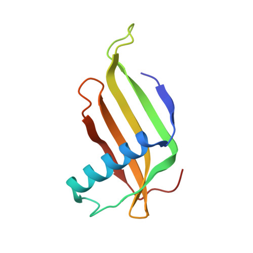
5O7K, 5O7L, 5O7Q, 5O7R, 5O7S - PubMed Abstract:
MNEI and its variant Y65R-MNEI are sweet proteins with potential applications as sweeteners in food industry. Also, they are often used as model systems for folding and aggregation studies. X-ray crystallography was used to structurally characterize Y65R-MNEI at five different pHs, while circular dichroism and fluorescence spectroscopy were used to study their thermal and chemical stability. ThT assay and AFM were used for studying the kinetics of aggregation and morphology of the aggregates. Crystal structures of Y65R-MNEI revealed the existence of a dimer in the asymmetric unit, which, depending on the pH, assumes either an open or a closed conformation. The pH dramatically affects kinetics of formation and morphology of the aggregates: both MNEI and Y65R-MNEI form fibrils at acidic pH while amorphous aggregates are observed at neutral pH. The mutation Y65R induces structural modifications at the C-terminal region of the protein, which account for the decreased stability of the mutant when compared to MNEI. Furthermore, the pH-dependent conformation of the Y65R-MNEI dimer may explain the different type of aggregates formed as a function of pH. The investigation of the structural bases of aggregation gets us closer to the possibility of controlling such process, either by tuning the physicochemical environmental parameters or by site directed mutagenesis. This knowledge is helpful to expand the range of stability of proteins with potential industrial applications, such as MNEI and its mutant Y65R-MNEI, which should ideally preserve their structure and soluble state through a wide array of conditions.
- Department of Chemical Sciences, University of Naples Federico II, Complesso Universitario di Monte Sant'Angelo, Via Cintia, I-80126 Napoli, Italy.
Organizational Affiliation: