5O79

Klebsiella pneumoniae OmpK36


Experimental Data Snapshot

  • Method: X-RAY DIFFRACTION
  • Resolution: 1.65 Å
  • R-Value Free: 0.190 
  • R-Value Work: 0.146 
  • R-Value Observed: 0.148 

wwPDB Validation   3D Report Full Report


Ligand Structure Quality Assessment 


This is version 1.2 of the entry. See complete history


Literature

Getting Drugs into Gram-Negative Bacteria: Rational Rules for Permeation through General Porins.

Acosta-Gutierrez, S.Ferrara, L.Pathania, M.Masi, M.Wang, J.Bodrenko, I.Zahn, M.Winterhalter, M.Stavenger, R.A.Pages, J.M.Naismith, J.H.van den Berg, B.Page, M.G.P.Ceccarelli, M.

(2018) ACS Infect Dis 4: 1487-1498

  • DOI: https://doi.org/10.1021/acsinfecdis.8b00108
  • Primary Citation of Related Structures:  
    5O77, 5O79, 6ENE

  • PubMed Abstract: 

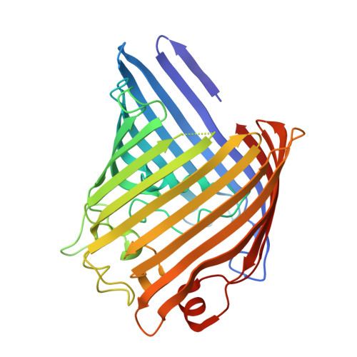
    Small, hydrophilic molecules, including most important antibiotics in clinical use, cross the Gram-negative outer membrane through the water-filled channels provided by porins. We have determined the X-ray crystal structures of the principal general porins from three species of Enterobacteriaceae, namely Enterobacter aerogenes, Enterobacter cloacae, and Klebsiella pneumoniae, and determined their antibiotic permeabilities as well as those of the orthologues from Escherichia coli. Starting from the structure of the porins and molecules, we propose a physical mechanism underlying transport and condense it in a computationally efficient scoring function. The scoring function shows good agreement with in vitro penetration data and will enable the screening of virtual databases to identify molecules with optimal permeability through porins and help to guide the optimization of antibiotics with poor permeation.


  • Organizational Affiliation

    Department of Physics , University of Cagliari, Cittadella Universitaria di Monserrato , SP Monserrato-Sestu Km 0.8 , Monserrato , 09042 , Italy.


Macromolecules
Find similar proteins by:  (by identity cutoff)  |  3D Structure
Entity ID: 1
MoleculeChains Sequence LengthOrganismDetailsImage
OmpK36
A, B, C
344Klebsiella pneumoniaeMutation(s): 0 
Gene Names: ompK36
Membrane Entity: Yes 
UniProt
Find proteins for D6QLY0 (Klebsiella pneumoniae)
Explore D6QLY0 
Go to UniProtKB:  D6QLY0
Entity Groups  
Sequence Clusters30% Identity50% Identity70% Identity90% Identity95% Identity100% Identity
UniProt GroupD6QLY0
Sequence Annotations
Expand
  • Reference Sequence
Small Molecules
Ligands 2 Unique
IDChains Name / Formula / InChI Key2D Diagram3D Interactions
C8E
Query on C8E

Download Ideal Coordinates CCD File 
D [auth A]
F [auth B]
G [auth B]
H [auth B]
I [auth B]
D [auth A],
F [auth B],
G [auth B],
H [auth B],
I [auth B],
J [auth B],
K [auth B],
M [auth C],
N [auth C]
(HYDROXYETHYLOXY)TRI(ETHYLOXY)OCTANE
C16 H34 O5
FEOZZFHAVXYAMB-UHFFFAOYSA-N
MG
Query on MG

Download Ideal Coordinates CCD File 
E [auth A],
L [auth B],
O [auth C]
MAGNESIUM ION
Mg
JLVVSXFLKOJNIY-UHFFFAOYSA-N
Experimental Data & Validation

Experimental Data

  • Method: X-RAY DIFFRACTION
  • Resolution: 1.65 Å
  • R-Value Free: 0.190 
  • R-Value Work: 0.146 
  • R-Value Observed: 0.148 
  • Space Group: C 1 2 1
Unit Cell:
Length ( Å )Angle ( ˚ )
a = 232.316α = 90
b = 74.511β = 112.18
c = 90.814γ = 90
Software Package:
Software NamePurpose
REFMACrefinement
XDSdata reduction
Aimlessdata scaling
MOLREPphasing

Structure Validation

View Full Validation Report


Ligand Structure Quality Assessment 


Entry History & Funding Information

Deposition Data


Funding OrganizationLocationGrant Number
European UnionUnited Kingdom--

Revision History  (Full details and data files)

  • Version 1.0: 2018-06-20
    Type: Initial release
  • Version 1.1: 2019-11-13
    Changes: Data collection, Database references
  • Version 1.2: 2024-01-17
    Changes: Data collection, Database references, Derived calculations, Refinement description