5O6G | pdb_00005o6g

Structures and dynamics of mesophilic variants from the homing endonuclease I-DmoI


Experimental Data Snapshot

  • Method: X-RAY DIFFRACTION
  • Resolution: 2.75 Å
  • R-Value Free: 
    0.232 (Depositor), 0.232 (DCC) 
  • R-Value Work: 
    0.177 (Depositor), 0.178 (DCC) 
  • R-Value Observed: 
    0.180 (Depositor) 

Starting Model: experimental
View more details

wwPDB Validation 3D Report Full Report

Validation slider image for 5O6G

This is version 1.3 of the entry. See complete history

Literature

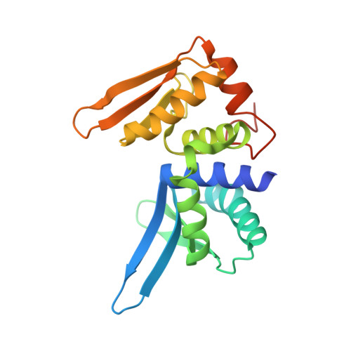
Structure and dynamics of mesophilic variants from the homing endonuclease I-DmoI.

Alba, J.Marcaida, M.J.Prieto, J.Montoya, G.Molina, R.D'Abramo, M.

(2017) J Comput Aided Mol Des 31: 1063-1072

  • DOI: https://doi.org/10.1007/s10822-017-0087-5
  • Primary Citation Related Structures: 
    5O6G, 5O6I

  • PubMed Abstract: 

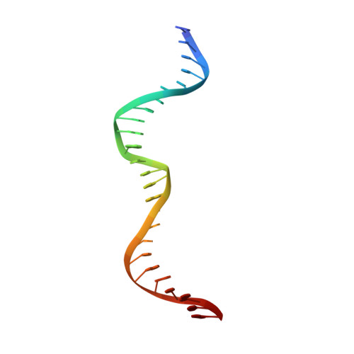
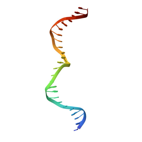
    I-DmoI, from the hyperthermophilic archaeon Desulfurococcus mobilis, belongs to the LAGLIDADG homing endonuclease protein family. Its members are highly specific enzymes capable of recognizing long DNA target sequences, thus providing potential tools for genome manipulation. Working towards this particular application, many efforts have been made to generate mesophilic variants of I-DmoI that function at lower temperatures than the wild-type. Here, we report a structural and computational analysis of two I-DmoI mesophilic mutants. Despite very limited structural variations between the crystal structures of these variants and the wild-type, a different dynamical behaviour near the cleavage sites is observed. In particular, both the dynamics of the water molecules and the protein perturbation effect on the cleavage site correlate well with the changes observed in the experimental enzymatic activity.


  • Organizational Affiliation
    • Department of Chemistry, Sapienza University of Rome, P.le A. Moro, 5, 00185, Rome, Italy.

Macromolecule Content 

  • Total Structure Weight: 116.79 kDa 
  • Atom Count: 7,676 
  • Modeled Residue Count: 694 
  • Deposited Residue Count: 750 
  • Unique protein chains: 1
  • Unique nucleic acid chains: 2

Macromolecules


Find similar proteins by:|  3D Structure
Entity ID: 1
MoleculeChains  Sequence LengthOrganismDetailsImage
Homing endonuclease I-DmoI
A, D, G
200Desulfurococcus mucosusMutation(s): 0 
EC: 3.1
UniProt
Find proteins for P21505 (Desulfurococcus mucosus)
Explore P21505 
Go to UniProtKB:  P21505
Entity Groups
Sequence Clusters30% Identity50% Identity70% Identity90% Identity95% Identity100% Identity
UniProt GroupP21505
Sequence Annotations
Expand
Reference Sequence
Find similar nucleic acids by:  Sequence
Entity ID: 2
MoleculeChains LengthOrganismImage
DNA (25-MER)
B, E, H
25synthetic construct
Sequence Annotations
Expand
Reference Sequence
Find similar nucleic acids by:  Sequence
Entity ID: 3
MoleculeChains LengthOrganismImage
DNA (25-MER)
C, F, I
25synthetic construct
Sequence Annotations
Expand
Reference Sequence

Experimental Data & Validation

Experimental Data

  • Method: X-RAY DIFFRACTION
  • Resolution: 2.75 Å
  • R-Value Free:  0.232 (Depositor), 0.232 (DCC) 
  • R-Value Work:  0.177 (Depositor), 0.178 (DCC) 
  • R-Value Observed: 0.180 (Depositor) 
Space Group: P 1 21 1
Unit Cell:
Length ( Å )Angle ( ˚ )
a = 107.28α = 90
b = 70.5β = 119.89
c = 107.15γ = 90
Software Package:
Software NamePurpose
PHENIXrefinement
XDSdata reduction
Aimlessdata scaling
PHASERphasing

Structure Validation

View Full Validation Report


Entry History 

Deposition Data

Revision History  (Full details and data files)

  • Version 1.0: 2017-12-06
    Type: Initial release
  • Version 1.1: 2018-01-03
    Changes: Database references
  • Version 1.2: 2019-10-16
    Changes: Data collection, Database references
  • Version 1.3: 2024-01-17
    Changes: Advisory, Data collection, Database references, Derived calculations, Refinement description