


Crystal structure determination from picosecond infrared laser ablated protein crystals by serial synchrotron crystallography
Schulz, E.C., Mehrabi, P., Pai, E.F., Miller, R.J.D.To be published.
Experimental Data Snapshot
wwPDB Validation 3D Report Full Report

Entity ID: 1 | |||||
|---|---|---|---|---|---|
| Molecule | Chains | Sequence Length | Organism | Details | Image |
| Fluoroacetate dehalogenase | 306 | Rhodopseudomonas palustris | Mutation(s): 0 Gene Names: RPA1163 EC: 3.8.1.3 | 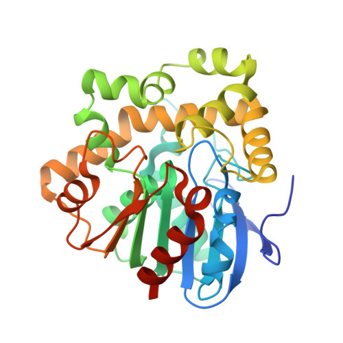 | |
UniProt | |||||
Entity Groups | |||||
| Sequence Clusters | 30% Identity50% Identity70% Identity90% Identity95% Identity100% Identity | ||||
| UniProt Group | Q6NAM1 | ||||
Sequence AnnotationsExpand | |||||
Reference Sequence | |||||
| Length ( Å ) | Angle ( ˚ ) |
|---|---|
| a = 42.413 | α = 90 |
| b = 80.349 | β = 102.96 |
| c = 85.648 | γ = 90 |
| Software Name | Purpose |
|---|---|
| PHENIX | refinement |
| cctbx.xfel | data reduction |
| cctbx.prime | data scaling |
| PHASER | phasing |


