5O1L

Structure of Latex Clearing Protein LCP in the open state with bound imidazole


Experimental Data Snapshot

  • Method: X-RAY DIFFRACTION
  • Resolution: 1.48 Å
  • R-Value Free: 0.189 
  • R-Value Work: 0.166 
  • R-Value Observed: 0.167 

wwPDB Validation   3D Report Full Report


Ligand Structure Quality Assessment 


This is version 1.0 of the entry. See complete history


Literature

Structural and Functional Analysis of Latex Clearing Protein (Lcp) Provides Insight into the Enzymatic Cleavage of Rubber.

Ilcu, L.Rother, W.Birke, J.Brausemann, A.Einsle, O.Jendrossek, D.

(2017) Sci Rep 7: 6179-6179

  • DOI: https://doi.org/10.1038/s41598-017-05268-2
  • Primary Citation of Related Structures:  
    5O1L, 5O1M

  • PubMed Abstract: 

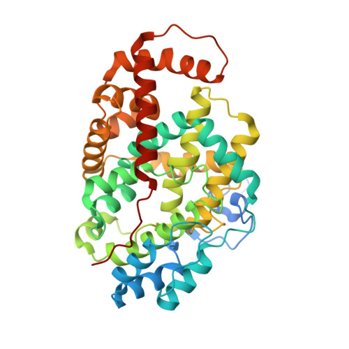
    Latex clearing proteins (Lcps) are rubber oxygenases that catalyse the extracellular cleavage of poly (cis-1,4-isoprene) by Gram-positive rubber degrading bacteria. Lcp of Streptomyces sp. K30 (Lcp K30 ) is a b-type cytochrome and acts as an endo-type dioxygenase producing C 20 and higher oligo-isoprenoids that differ in the number of isoprene units but have the same terminal functions, CHO-CH 2 - and -CH 2 -COCH 3 . Our analysis of the Lcp K30 structure revealed a 3/3 globin fold with additional domains at the N- and C-termini and similarities to globin-coupled sensor proteins. The haem group of Lcp K30 is ligated to the polypeptide by a proximal histidine (His198) and by a lysine residue (Lys167) as the distal axial ligand. The comparison of Lcp K30 structures in a closed and in an open state as well as spectroscopic and biochemical analysis of wild type and Lcp K30 muteins provided insights into the action of the enzyme during catalysis.


  • Organizational Affiliation

    Institute for Biochemistry, Albert-Ludwigs-Universität Freiburg, Albertstrasse 21, 79104, Freiburg, Germany.


Macromolecules
Find similar proteins by:  (by identity cutoff)  |  3D Structure
Entity ID: 1
MoleculeChains Sequence LengthOrganismDetailsImage
Rubber oxygenase
A, B
407Streptomyces sp. K30Mutation(s): 0 
Gene Names: lcp
EC: 1.13
UniProt
Find proteins for Q3L8N0 (Streptomyces sp. (strain K30))
Explore Q3L8N0 
Go to UniProtKB:  Q3L8N0
Entity Groups  
Sequence Clusters30% Identity50% Identity70% Identity90% Identity95% Identity100% Identity
UniProt GroupQ3L8N0
Sequence Annotations
Expand
  • Reference Sequence
Small Molecules
Ligands 4 Unique
IDChains Name / Formula / InChI Key2D Diagram3D Interactions
HEM
Query on HEM

Download Ideal Coordinates CCD File 
C [auth A],
H [auth B]
PROTOPORPHYRIN IX CONTAINING FE
C34 H32 Fe N4 O4
KABFMIBPWCXCRK-RGGAHWMASA-L
BU3
Query on BU3

Download Ideal Coordinates CCD File 
F [auth A],
K [auth B]
(R,R)-2,3-BUTANEDIOL
C4 H10 O2
OWBTYPJTUOEWEK-QWWZWVQMSA-N
IMD
Query on IMD

Download Ideal Coordinates CCD File 
D [auth A],
E [auth A],
I [auth B],
J [auth B]
IMIDAZOLE
C3 H5 N2
RAXXELZNTBOGNW-UHFFFAOYSA-O
EDO
Query on EDO

Download Ideal Coordinates CCD File 
G [auth A],
L [auth B]
1,2-ETHANEDIOL
C2 H6 O2
LYCAIKOWRPUZTN-UHFFFAOYSA-N
Experimental Data & Validation

Experimental Data

  • Method: X-RAY DIFFRACTION
  • Resolution: 1.48 Å
  • R-Value Free: 0.189 
  • R-Value Work: 0.166 
  • R-Value Observed: 0.167 
  • Space Group: P 1
Unit Cell:
Length ( Å )Angle ( ˚ )
a = 56.758α = 85.43
b = 62.796β = 66.06
c = 64.416γ = 74.16
Software Package:
Software NamePurpose
REFMACrefinement
XDSdata reduction
Aimlessdata scaling
PHASERphasing

Structure Validation

View Full Validation Report


Ligand Structure Quality Assessment 


Entry History & Funding Information

Deposition Data


Funding OrganizationLocationGrant Number
German Research FoundationGermanyEi-520/5
German Research FoundationGermanyJe-152/18

Revision History  (Full details and data files)

  • Version 1.0: 2017-08-02
    Type: Initial release