5O12 | pdb_00005o12

Crystal structure of PIM1 kinase in complex with small-molecule inhibitor


Experimental Data Snapshot

  • Method: X-RAY DIFFRACTION
  • Resolution: 2.40 Å
  • R-Value Free: 
    0.223 (Depositor), 0.212 (DCC) 
  • R-Value Work: 
    0.177 (Depositor) 
  • R-Value Observed: 
    0.179 (Depositor) 

Starting Model: experimental
View more details

wwPDB Validation 3D Report Full Report

Validation slider image for 5O12

Ligand Structure Quality Assessment 


This is version 1.3 of the entry. See complete history

Literature

Structural analysis of PIM1 kinase complexes with ATP-competitive inhibitors.

Bogusz, J.Zrubek, K.Rembacz, K.P.Grudnik, P.Golik, P.Romanowska, M.Wladyka, B.Dubin, G.

(2017) Sci Rep 7: 13399-13399

  • DOI: https://doi.org/10.1038/s41598-017-13557-z
  • Primary Citation Related Structures: 
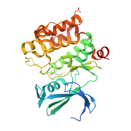
    5O11, 5O12, 5O13

  • PubMed Abstract: 

    PIM1 is an oncogenic kinase overexpressed in a number of cancers where it correlates with poor prognosis. Several studies demonstrated that inhibition of PIM1 activity is an attractive strategy in fighting overexpressing cancers, while distinct structural features of ATP binding pocket make PIM1 an inviting target for the design of selective inhibitors. To facilitate development of specific PIM1 inhibitors, in this study we report three crystal structures of ATP-competitive inhibitors at the ATP binding pocket of PIM1. Two of the reported structures (CX-4945 and Ro-3306) explain the off-target effect on PIM1 of respectively casein kinase 2 and cyclin-dependent kinase 1 dedicated inhibitors. In turn, the structure with CX-6258 demonstrates a binding mode of a potent, selective inhibitor of PIM1, PIM2, PIM3 and Flt-3 kinases. The consequences of our findings for future inhibitor development are discussed.


  • Organizational Affiliation
    • Malopolska Centre of Biotechnology, Jagiellonian University, Gronostajowa 7a, 30-387, Krakow, Poland.

Macromolecule Content 

  • Total Structure Weight: 34.95 kDa 
  • Atom Count: 2,211 
  • Modeled Residue Count: 266 
  • Deposited Residue Count: 298 
  • Unique protein chains: 1

Macromolecules

Find similar proteins by:|  3D Structure
Entity ID: 1
MoleculeChains  Sequence LengthOrganismDetailsImage
Serine/threonine-protein kinase pim-1298Homo sapiensMutation(s): 0 
Gene Names: PIM1
EC: 2.7.11.1
UniProt & NIH Common Fund Data Resources
Find proteins for P11309 (Homo sapiens)
Explore P11309 
Go to UniProtKB:  P11309
PHAROS:  P11309
GTEx:  ENSG00000137193 
Entity Groups
Sequence Clusters30% Identity50% Identity70% Identity90% Identity95% Identity100% Identity
UniProt GroupP11309
Sequence Annotations
Expand
Reference Sequence

Small Molecules

Ligands 2 Unique
IDChains Name / Formula / InChI Key2D Diagram3D Interactions
1RO

Query on 1RO


Download:Ideal Coordinates CCD File
B [auth A](5E)-5-(quinolin-6-ylmethylidene)-2-[(thiophen-2-ylmethyl)amino]-1,3-thiazol-4(5H)-one
C18 H13 N3 O S2
XOLMRFUGOINFDQ-MHWRWJLKSA-N
IMD

Query on IMD


Download:Ideal Coordinates CCD File
C [auth A]IMIDAZOLE
C3 H5 N2
RAXXELZNTBOGNW-UHFFFAOYSA-O
Modified Residues  2 Unique
IDChains TypeFormula2D DiagramParent
CME
Query on CME
A
L-PEPTIDE LINKINGC5 H11 N O3 S2CYS
SEP
Query on SEP
A
L-PEPTIDE LINKINGC3 H8 N O6 PSER

Experimental Data & Validation

Experimental Data

  • Method: X-RAY DIFFRACTION
  • Resolution: 2.40 Å
  • R-Value Free:  0.223 (Depositor), 0.212 (DCC) 
  • R-Value Work:  0.177 (Depositor) 
  • R-Value Observed: 0.179 (Depositor) 
Space Group: P 65
Unit Cell:
Length ( Å )Angle ( ˚ )
a = 97.672α = 90
b = 97.672β = 90
c = 80.579γ = 120
Software Package:
Software NamePurpose
REFMACrefinement
XDSdata reduction
SCALAdata scaling
PHASERphasing

Structure Validation

View Full Validation Report


Ligand Structure Quality Assessment 


Entry History 

& Funding Information

Deposition Data


Funding OrganizationLocationGrant Number
National Science CenterPolandUMO-2012/07/E/NZ1/01907

Revision History  (Full details and data files)

  • Version 1.0: 2017-11-01
    Type: Initial release
  • Version 1.1: 2019-10-16
    Changes: Data collection
  • Version 1.2: 2024-01-17
    Changes: Data collection, Database references, Refinement description
  • Version 1.3: 2024-10-16
    Changes: Structure summary