5O09

BtubABC mini microtubule


Experimental Data Snapshot

  • Method: ELECTRON MICROSCOPY
  • Resolution: 3.60 Å
  • Aggregation State: HELICAL ARRAY 
  • Reconstruction Method: HELICAL 

wwPDB Validation   3D Report Full Report


This is version 1.4 of the entry. See complete history


Literature

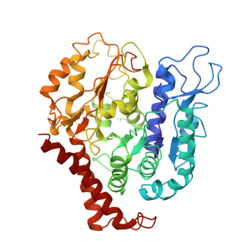
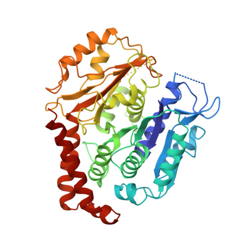
Four-stranded mini microtubules formed by Prosthecobacter BtubAB show dynamic instability.

Deng, X.Fink, G.Bharat, T.A.M.He, S.Kureisaite-Ciziene, D.Lowe, J.

(2017) Proc Natl Acad Sci U S A 114: E5950-E5958

  • DOI: https://doi.org/10.1073/pnas.1705062114
  • Primary Citation of Related Structures:  
    5O01, 5O09

  • PubMed Abstract: 

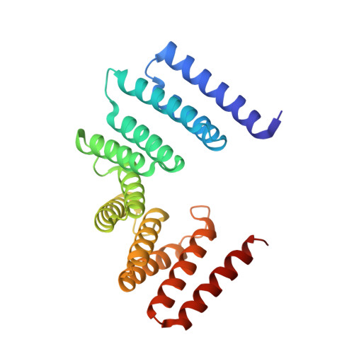
    Microtubules, the dynamic, yet stiff hollow tubes built from αβ-tubulin protein heterodimers, are thought to be present only in eukaryotic cells. Here, we report a 3.6-Å helical reconstruction electron cryomicroscopy structure of four-stranded mini microtubules formed by bacterial tubulin-like Prosthecobacter dejongeii BtubAB proteins. Despite their much smaller diameter, mini microtubules share many key structural features with eukaryotic microtubules, such as an M-loop, alternating subunits, and a seam that breaks overall helical symmetry. Using in vitro total internal reflection fluorescence microscopy, we show that bacterial mini microtubules treadmill and display dynamic instability, another hallmark of eukaryotic microtubules. The third protein in the btub gene cluster, BtubC, previously known as "bacterial kinesin light chain," binds along protofilaments every 8 nm, inhibits BtubAB mini microtubule catastrophe, and increases rescue. Our work reveals that some bacteria contain regulated and dynamic cytomotive microtubule systems that were once thought to be only useful in much larger and sophisticated eukaryotic cells.


  • Organizational Affiliation

    Structural Studies Division, Medical Research Council Laboratory of Molecular Biology, Cambridge CB2 0QH, United Kingdom.


Macromolecules
Find similar proteins by:  (by identity cutoff)  |  3D Structure
Entity ID: 1
MoleculeChains Sequence LengthOrganismDetailsImage
Tubulin433Prosthecobacter dejongeiiMutation(s): 0 
Gene Names: btubA
UniProt
Find proteins for Q8GCC5 (Prosthecobacter dejongeii)
Explore Q8GCC5 
Go to UniProtKB:  Q8GCC5
Entity Groups  
Sequence Clusters30% Identity50% Identity70% Identity90% Identity95% Identity100% Identity
UniProt GroupQ8GCC5
Sequence Annotations
Expand
  • Reference Sequence
Find similar proteins by:  (by identity cutoff)  |  3D Structure
Entity ID: 2
MoleculeChains Sequence LengthOrganismDetailsImage
Tubulin BtubB426Prosthecobacter dejongeiiMutation(s): 0 
Gene Names: btubB
UniProt
Find proteins for Q8GCC1 (Prosthecobacter dejongeii)
Explore Q8GCC1 
Go to UniProtKB:  Q8GCC1
Entity Groups  
Sequence Clusters30% Identity50% Identity70% Identity90% Identity95% Identity100% Identity
UniProt GroupQ8GCC1
Sequence Annotations
Expand
  • Reference Sequence
Find similar proteins by:  (by identity cutoff)  |  3D Structure
Entity ID: 3
MoleculeChains Sequence LengthOrganismDetailsImage
Bacterial kinesin light chain238Prosthecobacter vanneerveniiMutation(s): 0 
Gene Names: bklc
UniProt
Find proteins for A8Y5U5 (Prosthecobacter vanneervenii)
Explore A8Y5U5 
Go to UniProtKB:  A8Y5U5
Entity Groups  
Sequence Clusters30% Identity50% Identity70% Identity90% Identity95% Identity100% Identity
UniProt GroupA8Y5U5
Sequence Annotations
Expand
  • Reference Sequence
Small Molecules
Ligands 1 Unique
IDChains Name / Formula / InChI Key2D Diagram3D Interactions
GDP
Query on GDP

Download Ideal Coordinates CCD File 
AA [auth 2A]
BA [auth 2B]
CA [auth 3A]
DA [auth 3B]
EA [auth 4A]
AA [auth 2A],
BA [auth 2B],
CA [auth 3A],
DA [auth 3B],
EA [auth 4A],
FA [auth 4B],
GA [auth 5A],
HA [auth 5B],
IA [auth 6A],
JA [auth 6B],
KA [auth 7A],
LA [auth 7B],
MA [auth 8A],
NA [auth 8B],
Y [auth 1A],
Z [auth 1B]
GUANOSINE-5'-DIPHOSPHATE
C10 H15 N5 O11 P2
QGWNDRXFNXRZMB-UUOKFMHZSA-N
Experimental Data & Validation

Experimental Data

  • Method: ELECTRON MICROSCOPY
  • Resolution: 3.60 Å
  • Aggregation State: HELICAL ARRAY 
  • Reconstruction Method: HELICAL 

Structure Validation

View Full Validation Report


Entry History 

Deposition Data

Revision History  (Full details and data files)

  • Version 1.0: 2017-07-19
    Type: Initial release
  • Version 1.1: 2017-07-26
    Changes: Database references
  • Version 1.2: 2017-08-30
    Changes: Refinement description
  • Version 1.3: 2017-10-04
    Changes: Other
  • Version 1.4: 2018-10-03
    Changes: Advisory, Data collection, Derived calculations