On the (un)coupling of the chromophore, tongue interactions, and overall conformation in a bacterial phytochrome.
Takala, H., Lehtivuori, H.K., Berntsson, O., Hughes, A., Nanekar, R., Niebling, S., Panman, M., Henry, L., Menzel, A., Westenhoff, S., Ihalainen, J.A.(2018) J Biol Chem 293: 8161-8172
- PubMed: 29622676 Search on PubMedSearch on PubMed Central
- DOI: https://doi.org/10.1074/jbc.RA118.001794
- Primary Citation Related Structures:
5NFX, 5NM3, 5NWN - PubMed Abstract:
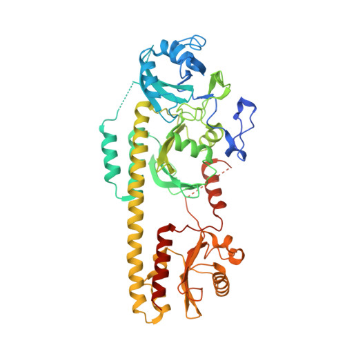
Phytochromes are photoreceptors in plants, fungi, and various microorganisms and cycle between metastable red light-absorbing (Pr) and far-red light-absorbing (Pfr) states. Their light responses are thought to follow a conserved structural mechanism that is triggered by isomerization of the chromophore. Downstream structural changes involve refolding of the so-called tongue extension of the phytochrome-specific GAF-related (PHY) domain of the photoreceptor. The tongue is connected to the chromophore by conserved DIP and PR X SF motifs and a conserved tyrosine, but the role of these residues in signal transduction is not clear. Here, we examine the tongue interactions and their interplay with the chromophore by substituting the conserved tyrosine (Tyr 263 ) in the phytochrome from the extremophile bacterium Deinococcus radiodurans with phenylalanine. Using optical and FTIR spectroscopy, X-ray solution scattering, and crystallography of chromophore-binding domain (CBD) and CBD-PHY fragments, we show that the absence of the Tyr 263 hydroxyl destabilizes the β-sheet conformation of the tongue. This allowed the phytochrome to adopt an α-helical tongue conformation regardless of the chromophore state, hence distorting the activity state of the protein. Our crystal structures further revealed that water interactions are missing in the Y263F mutant, correlating with a decrease of the photoconversion yield and underpinning the functional role of Tyr 263 in phytochrome conformational changes. We propose a model in which isomerization of the chromophore, refolding of the tongue, and globular conformational changes are represented as weakly coupled equilibria. The results also suggest that the phytochromes have several redundant signaling routes.
- Anatomy Department, Faculty of Medicine, University of Helsinki, Helsinki FI-00014, Finland; Departments of Biological and Environmental Sciences, University of Jyvaskyla, Jyvaskyla FI-40014, Finland. Electronic address: heikki.takala@helsinki.fi.
Organizational Affiliation: