5NVG | pdb_00005nvg

Thr12 Phosphorylated Ubiquitin


Experimental Data Snapshot

  • Method: X-RAY DIFFRACTION
  • Resolution: 1.07 Å
  • R-Value Free: 
    0.167 (Depositor), 0.131 (DCC) 
  • R-Value Work: 
    0.129 (Depositor), 0.131 (DCC) 
  • R-Value Observed: 
    0.131 (Depositor) 

wwPDB Validation 3D Report Full Report

Validation slider image for 5NVG

This is version 1.4 of the entry. See complete history

Literature

Biosynthesis and genetic encoding of phosphothreonine through parallel selection and deep sequencing.

Zhang, M.S.Brunner, S.F.Huguenin-Dezot, N.Liang, A.D.Schmied, W.H.Rogerson, D.T.Chin, J.W.

(2017) Nat Methods 14: 729-736

  • DOI: https://doi.org/10.1038/nmeth.4302
  • Primary Citation Related Structures: 
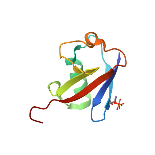
    5NVG

  • PubMed Abstract: 

    The phosphorylation of threonine residues in proteins regulates diverse processes in eukaryotic cells, and thousands of threonine phosphorylations have been identified. An understanding of how threonine phosphorylation regulates biological function will be accelerated by general methods to biosynthesize defined phosphoproteins. Here we describe a rapid approach for directly discovering aminoacyl-tRNA synthetase-tRNA pairs that selectively incorporate non-natural amino acids into proteins; our method uses parallel positive selections combined with deep sequencing and statistical analysis and enables the direct, scalable discovery of aminoacyl-tRNA synthetase-tRNA pairs with mutually orthogonal substrate specificity. By combining a method to biosynthesize phosphothreonine in cells with this selection approach, we discover a phosphothreonyl-tRNA synthetase-tRNA CUA pair and create an entirely biosynthetic route to incorporating phosphothreonine in proteins. We biosynthesize several phosphoproteins and demonstrate phosphoprotein structure determination and synthetic protein kinase activation.


  • Organizational Affiliation
    • Medical Research Council Laboratory of Molecular Biology, Francis Crick Avenue, Cambridge, England, UK.

Macromolecule Content 

  • Total Structure Weight: 8.86 kDa 
  • Atom Count: 825 
  • Modeled Residue Count: 76 
  • Deposited Residue Count: 76 
  • Unique protein chains: 1

Macromolecules

Find similar proteins by:|  3D Structure
Entity ID: 1
MoleculeChains  Sequence LengthOrganismDetailsImage
Polyubiquitin-B76Homo sapiensMutation(s): 0 
Gene Names: UBB
UniProt & NIH Common Fund Data Resources
Find proteins for P0CG47 (Homo sapiens)
Explore P0CG47 
Go to UniProtKB:  P0CG47
PHAROS:  P0CG47
GTEx:  ENSG00000170315 
Entity Groups
Sequence Clusters30% Identity50% Identity70% Identity90% Identity95% Identity100% Identity
UniProt GroupP0CG47
Sequence Annotations
Expand
Reference Sequence

Small Molecules

Modified Residues  1 Unique
IDChains TypeFormula2D DiagramParent
TPO
Query on TPO
A
L-PEPTIDE LINKINGC4 H10 N O6 PTHR

Experimental Data & Validation

Experimental Data

  • Method: X-RAY DIFFRACTION
  • Resolution: 1.07 Å
  • R-Value Free:  0.167 (Depositor), 0.131 (DCC) 
  • R-Value Work:  0.129 (Depositor), 0.131 (DCC) 
  • R-Value Observed: 0.131 (Depositor) 
Space Group: C 1 2 1
Unit Cell:
Length ( Å )Angle ( ˚ )
a = 50.46α = 90
b = 24.74β = 113.17
c = 47.65γ = 90
Software Package:
Software NamePurpose
REFMACrefinement
Cootmodel building
PHASERphasing
iMOSFLMdata reduction
Aimlessdata scaling

Structure Validation

View Full Validation Report


Entry History 

& Funding Information

Deposition Data


Funding OrganizationLocationGrant Number
Medical Research Council (United Kingdom)United KingdomMC_U105181009
Medical Research Council (United Kingdom)United KingdomMC_UP_A024_1008

Revision History  (Full details and data files)

  • Version 1.0: 2017-05-31
    Type: Initial release
  • Version 1.1: 2017-06-07
    Changes: Database references
  • Version 1.2: 2017-07-12
    Changes: Database references
  • Version 1.3: 2017-09-13
    Changes: Author supporting evidence
  • Version 1.4: 2024-10-23
    Changes: Data collection, Database references, Derived calculations, Structure summary