5NSJ | pdb_00005nsj

GP1 receptor-binding domain from Whitewater Arroyo mammarenavirus


Experimental Data Snapshot

  • Method: X-RAY DIFFRACTION
  • Resolution: 2.25 Å
  • R-Value Free: 
    0.209 (Depositor), 0.211 (DCC) 
  • R-Value Work: 
    0.183 (Depositor) 
  • R-Value Observed: 
    0.186 (Depositor) 

Starting Model: experimental
View more details

wwPDB Validation 3D Report Full Report

Validation slider image for 5NSJ

Ligand Structure Quality Assessment 


This is version 2.2 of the entry. See complete history

Literature

Structural Basis for Receptor Selectivity by the Whitewater Arroyo Mammarenavirus.

Shimon, A.Shani, O.Diskin, R.

(2017) J Mol Biology 429: 2825-2839

  • DOI: https://doi.org/10.1016/j.jmb.2017.07.011
  • Primary Citation Related Structures: 
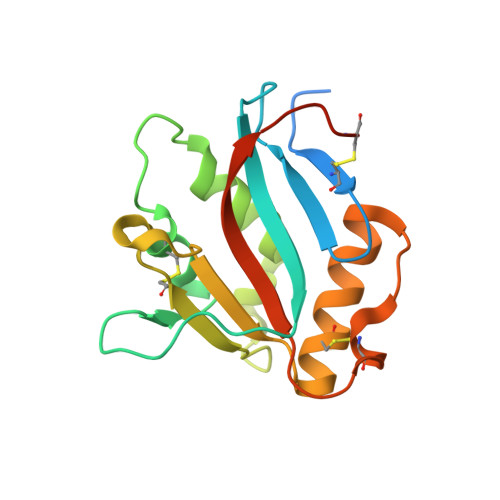
    5NSJ

  • PubMed Abstract: 

    Whitewater Arroyo virus belongs to the "New World" group of mammarenaviruses that reside in rodent reservoirs and are prevalent in North and South Americas. Clades B and A/B of New World mammarenaviruses use transferrin receptor 1 (TfR1) for entry. While all of these viruses use rodent-derived TfR1 orthologs, some can also use the human-TfR1 and thereby infect humans. Although we have structural information for TfR1 recognition by pathogenic virus, we do not know what the structural differences are between the receptor-binding domains of pathogenic and non-pathogenic viruses that allow some but not all viruses to utilize the human receptor for entry. The poor understanding of the molecular determinants of mammarenavirus host range, and thus pathogenicity, is partly due to the low sequence similarity between the receptor-binding domains from these viruses and the limited available structural information that preclude the use of modeling approaches. Here we present the first crystal structure of a receptor-binding domain of a non-pathogenic clade A/B mammarenavirus. This structure reveals the magnitude of structural differences within the receptor-binding domains of TfR1-tropic viruses. Our structural and sequence analyses indicate that the same structural incompatibilities with the human receptor equally affect both pathogenic and non-pathogenic mammarenaviruses. Non-pathogenic viruses do not have specific structural elements that prevent them from using the human receptor. Instead, the ability to utilize the human receptor directly depends on the extent of weak interactions throughout the receptor-binding site that in some viruses are sufficiently strong to overcome the structural incompatibilities.


  • Organizational Affiliation
    • Department of Structural Biology, Weizmann Institute of Science, Rehovot, Israel.

Macromolecule Content 

  • Total Structure Weight: 40.27 kDa 
  • Atom Count: 2,705 
  • Modeled Residue Count: 284 
  • Deposited Residue Count: 334 
  • Unique protein chains: 1

Macromolecules

Find similar proteins by:|  3D Structure
Entity ID: 1
MoleculeChains  Sequence LengthOrganismDetailsImage
Pre-glycoprotein polyprotein GP complex
A, B
167Mammarenavirus whitewaterenseMutation(s): 0 
Gene Names: GPCGP-C
UniProt
Find proteins for Q911P0 (Whitewater Arroyo mammarenavirus (isolate Rat/United States/AV 9310135/1995))
Explore Q911P0 
Go to UniProtKB:  Q911P0
Entity Groups
Sequence Clusters30% Identity50% Identity70% Identity90% Identity95% Identity100% Identity
UniProt GroupQ911P0
Glycosylation
Glycosylation Sites: 2
Sequence Annotations
Expand
Reference Sequence

Oligosaccharides

Help  
Entity ID: 2
MoleculeChains Length2D Diagram GlycosylationD Interactions
2-acetamido-2-deoxy-beta-D-glucopyranose-(1-4)-2-acetamido-2-deoxy-beta-D-glucopyranose
C, D
2N-Glycosylation
Glycosylation Resources
GlyTouCan: G42666HT
GlyCosmos: G42666HT
GlyGen: G42666HT
Entity ID: 3
MoleculeChains Length2D Diagram GlycosylationD Interactions
alpha-D-mannopyranose-(1-4)-2-acetamido-2-deoxy-beta-D-glucopyranose-(1-4)-2-acetamido-2-deoxy-beta-D-glucopyranose
E
3N-Glycosylation
Glycosylation Resources
GlyTouCan: G62182OO
GlyCosmos: G62182OO
GlyGen: G62182OO

Small Molecules

Ligands 2 Unique
IDChains Name / Formula / InChI Key2D Diagram3D Interactions
NAG

Query on NAG


Download:Ideal Coordinates CCD File
J [auth B]2-acetamido-2-deoxy-beta-D-glucopyranose
C8 H15 N O6
OVRNDRQMDRJTHS-FMDGEEDCSA-N
SM

Query on SM


Download:Ideal Coordinates CCD File
F [auth A]
G [auth A]
H [auth A]
I [auth A]
K [auth B]
F [auth A],
G [auth A],
H [auth A],
I [auth A],
K [auth B],
L [auth B],
M [auth B]
SAMARIUM (III) ION
Sm
DOSGOCSVHPUUIA-UHFFFAOYSA-N

Experimental Data & Validation

Experimental Data

  • Method: X-RAY DIFFRACTION
  • Resolution: 2.25 Å
  • R-Value Free:  0.209 (Depositor), 0.211 (DCC) 
  • R-Value Work:  0.183 (Depositor) 
  • R-Value Observed: 0.186 (Depositor) 
Space Group: C 2 2 21
Unit Cell:
Length ( Å )Angle ( ˚ )
a = 128.024α = 90
b = 128.352β = 90
c = 45.966γ = 90
Software Package:
Software NamePurpose
REFMACrefinement
HKL-2000data reduction
HKL-2000data scaling
AutoSolphasing
XDSdata reduction
Cootmodel building

Structure Validation

View Full Validation Report


Ligand Structure Quality Assessment 


Entry History 

& Funding Information

Deposition Data


Funding OrganizationLocationGrant Number
Israel Science FoundationIsrael682/16
I-COREIsrael1775/12

Revision History  (Full details and data files)

  • Version 1.0: 2017-08-02
    Type: Initial release
  • Version 1.1: 2017-09-06
    Changes: Database references, Refinement description, Structure summary
  • Version 2.0: 2020-07-29
    Type: Remediation
    Reason: Carbohydrate remediation
    Changes: Advisory, Atomic model, Data collection, Derived calculations, Structure summary
  • Version 2.1: 2024-01-17
    Changes: Data collection, Database references, Refinement description, Structure summary
  • Version 2.2: 2024-11-20
    Changes: Structure summary