5NS7

Crystal structure of beta-glucosidase BglM-G1 mutant H75R from marine metagenome


Experimental Data Snapshot

  • Method: X-RAY DIFFRACTION
  • Resolution: 1.54 Å
  • R-Value Free: 0.172 
  • R-Value Work: 0.159 
  • R-Value Observed: 0.159 

wwPDB Validation   3D Report Full Report


This is version 1.1 of the entry. See complete history


Literature

Loss of a conserved salt bridge in bacterial glycosyl hydrolase BgIM-G1 improves substrate binding in temperate environments.

Mhaindarkar, D.Gasper, R.Lupilov, N.Hofmann, E.Leichert, L.I.

(2018) Commun Biol 1: 171-171

  • DOI: https://doi.org/10.1038/s42003-018-0167-7
  • Primary Citation of Related Structures:  
    5NS6, 5NS7, 5NS8

  • PubMed Abstract: 

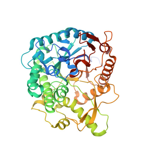
    Salt bridges are the strongest electrostatic interactions in proteins. They substantially contribute to a protein's structural stability. Thus, mutations of salt bridges are typically selected against. Here, we report on the evolutionary loss of a highly conserved salt bridge in the GH1 family glycosyl hydrolase BglM-G1. BglM-G1's gene was found in the bacterial metagenome of a temperate, seasonally cold marine habitat. In BglM-G1, arginine 75 is replaced by a histidine. While fully retaining β-glucosidase activity, BglM-G1 is less heat stable than an H75R variant, in which the salt bridge was artificially re-introduced. However, the K m toward its substrates was lower in wild type, leading to an overall higher catalytic efficiency. Our results indicate that this loss of the salt bridge leads to higher flexibility in BglM-G1's active site, trading structural stability at high temperatures, a trait not needed in a temperate, seasonally cold habitat, for a more effective catalytic activity.


  • Organizational Affiliation

    Ruhr University Bochum, Fakultät für Medizin, Institute for Biochemistry and Pathobiochemistry, Microbial Biochemistry, Universitätsstr. 150, 44780, Bochum, Germany.


Macromolecules
Find similar proteins by:  (by identity cutoff)  |  3D Structure
Entity ID: 1
MoleculeChains Sequence LengthOrganismDetailsImage
beta-glucosidase M - G1
A, B, C
454marine metagenomeMutation(s): 0 
EC: 3.2.1.21
Entity Groups  
Sequence Clusters30% Identity50% Identity70% Identity90% Identity95% Identity100% Identity
Sequence Annotations
Expand
  • Reference Sequence
Small Molecules
Ligands 2 Unique
IDChains Name / Formula / InChI Key2D Diagram3D Interactions
SO4
Query on SO4

Download Ideal Coordinates CCD File 
D [auth A]
E [auth A]
K [auth B]
L [auth B]
M [auth B]
D [auth A],
E [auth A],
K [auth B],
L [auth B],
M [auth B],
U [auth C],
V [auth C],
W [auth C]
SULFATE ION
O4 S
QAOWNCQODCNURD-UHFFFAOYSA-L
GOL
Query on GOL

Download Ideal Coordinates CCD File 
AA [auth C]
BA [auth C]
F [auth A]
G [auth A]
H [auth A]
AA [auth C],
BA [auth C],
F [auth A],
G [auth A],
H [auth A],
I [auth A],
J [auth A],
N [auth B],
O [auth B],
P [auth B],
Q [auth B],
R [auth B],
S [auth B],
T [auth B],
X [auth C],
Y [auth C],
Z [auth C]
GLYCEROL
C3 H8 O3
PEDCQBHIVMGVHV-UHFFFAOYSA-N
Experimental Data & Validation

Experimental Data

  • Method: X-RAY DIFFRACTION
  • Resolution: 1.54 Å
  • R-Value Free: 0.172 
  • R-Value Work: 0.159 
  • R-Value Observed: 0.159 
  • Space Group: P 43
Unit Cell:
Length ( Å )Angle ( ˚ )
a = 137.92α = 90
b = 137.92β = 90
c = 98.04γ = 90
Software Package:
Software NamePurpose
XSCALEdata scaling
PHENIXrefinement
PDB_EXTRACTdata extraction
XDSdata reduction

Structure Validation

View Full Validation Report


Entry History & Funding Information

Deposition Data


Funding OrganizationLocationGrant Number
European Research CouncilGermany281384

Revision History  (Full details and data files)

  • Version 1.0: 2018-08-08
    Type: Initial release
  • Version 1.1: 2019-01-30
    Changes: Data collection, Database references, Structure summary