5NPY | pdb_00005npy

Crystal structure of Helicobacter pylori flagellar hook protein FlgE2


Experimental Data Snapshot

  • Method: X-RAY DIFFRACTION
  • Resolution: 2.29 Å
  • R-Value Free: 
    0.266 (Depositor), 0.265 (DCC) 
  • R-Value Work: 
    0.188 (Depositor), 0.190 (DCC) 
  • R-Value Observed: 
    0.192 (Depositor) 

wwPDB Validation 3D Report Full Report

Validation slider image for 5NPY

This is version 1.2 of the entry. See complete history

Literature

Structural characterization of FlgE2 protein from Helicobacter pylori hook.

Loconte, V.Kekez, I.Matkovic-Calogovic, D.Zanotti, G.

(2017) FEBS J 284: 4328-4342

  • DOI: https://doi.org/10.1111/febs.14312
  • Primary Citation Related Structures: 
    5NPY

  • PubMed Abstract: 

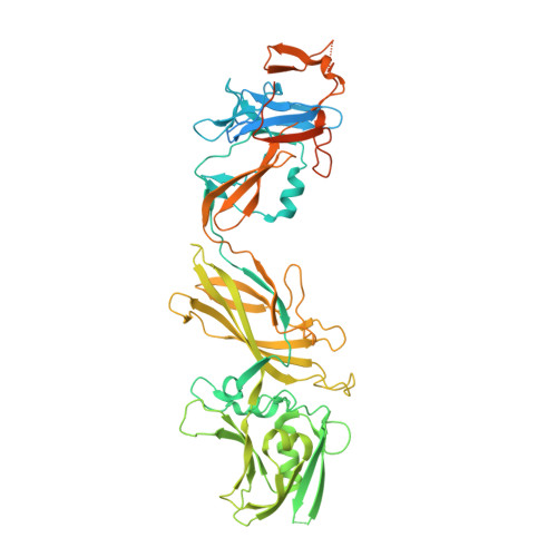
    The Helicobacter pylori flagellum is a complex rotatory nanomachine fundamental for the bacterium's survival in the human stomach. Protein FlgE is a component of the hook, a flexible junction exposed on the cell surface. In the H. pylori genome two different genes are present in different positions coding for hypothetical FlgE. The first protein, FlgE1, is the actual component of the flagellum hook, whilst the second, FlgE2, shares only 26% of the sequence identity with the other and its physiological function is still undefined. We have cloned, purified and crystallized FlgE2, whose structure, determined by the single-wavelength anomalous diffraction method, shows that in overall organization, the protein is composed of three distinct domains, two of them relatively similar to those of FlgE from other Gram-negative bacteria, whilst the third is peculiar to H. pylori. The crystal structure, along with the detected interaction with the regulatory cap protein FlgD, suggests a complementary function of FlgE1 and FlgE2 in the H. pylori flagellum, possibly typical of polar flagella, confirming the role of both proteins in the flagellar hook organization. Although some general features are shared with other Gram-negative bacteria, the presence of two different hook proteins indicates that the molecular organization of H. pylori flagellum has its own peculiarities. Atomic coordinates and structural factors have been deposited in the Protein Data Bank as 5NPY.


  • Organizational Affiliation
    • Department of Biomedical Sciences, University of Padua, Italy.

Macromolecule Content 

  • Total Structure Weight: 69.03 kDa 
  • Atom Count: 4,031 
  • Modeled Residue Count: 472 
  • Deposited Residue Count: 618 
  • Unique protein chains: 1

Macromolecules

Find similar proteins by:|  3D Structure
Entity ID: 1
MoleculeChains  Sequence LengthOrganismDetailsImage
Flagellar basal body protein618Helicobacter pylori G27Mutation(s): 0 
Gene Names: flgE-2HPG27_859
UniProt
Find proteins for B5Z7R4 (Helicobacter pylori (strain G27))
Explore B5Z7R4 
Go to UniProtKB:  B5Z7R4
Entity Groups
Sequence Clusters30% Identity50% Identity70% Identity90% Identity95% Identity100% Identity
UniProt GroupB5Z7R4
Sequence Annotations
Expand
Reference Sequence

Small Molecules

Ligands 1 Unique
IDChains Name / Formula / InChI Key2D Diagram3D Interactions
144

Query on 144


Download:Ideal Coordinates CCD File
B [auth A]TRIS-HYDROXYMETHYL-METHYL-AMMONIUM
C4 H12 N O3
DRDCQJADRSJFFD-UHFFFAOYSA-N

Experimental Data & Validation

Experimental Data

  • Method: X-RAY DIFFRACTION
  • Resolution: 2.29 Å
  • R-Value Free:  0.266 (Depositor), 0.265 (DCC) 
  • R-Value Work:  0.188 (Depositor), 0.190 (DCC) 
  • R-Value Observed: 0.192 (Depositor) 
Space Group: P 21 21 21
Unit Cell:
Length ( Å )Angle ( ˚ )
a = 43.209α = 90
b = 49.592β = 90
c = 238.261γ = 90
Software Package:
Software NamePurpose
PHENIXrefinement
iMOSFLMdata reduction
Aimlessdata scaling
autoSHARPphasing

Structure Validation

View Full Validation Report


Entry History 

Deposition Data

Revision History  (Full details and data files)

  • Version 1.0: 2017-11-15
    Type: Initial release
  • Version 1.1: 2017-12-27
    Changes: Database references
  • Version 1.2: 2024-05-08
    Changes: Data collection, Database references